
BIOLOGICAL COMPONENTS IDENTIFIED
copyright 2000 by Clifford E Carnicom
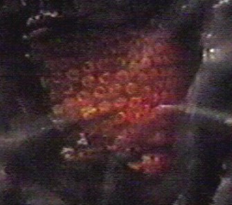
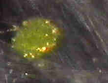
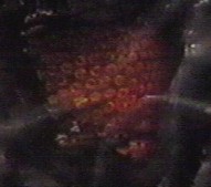
Biological components have now been identified in the two ground samples previously analyzed on www.carnicom.com. Numerous red blood cells, white blood cells, and unidentified cell types have been found within the sub-micron fiber sample previously presented and submitted on Jan 20 2000 to Carol M. Browner, Administrator of the United States Environmental Protection Agency. To date, Ms. Browner has refused to identify the sample delivered to her by certified mail, and to disclose those results to the American public. A visual analysis has now been conducted with a professional quality microscope on May 7 2000 that reveals the important discovery above. More information and images from this analysis will be presented in the future. Depicted above is one of two remarkable discoveries of clustered red blood cells which become readily visible after being subjected to immersion oil. The cells appear to be of a freeze-dried or dessicated nature in their original form within the microscopic fibers. Isolated and individual blood cells are interspersed throughout both of the samples which have previously been described. The surface of the cells appear to be modified in some way, but electron microscopy will likely be required to establish further detail. Professional medical analysis of the images and chemical analysis of the fibers, and the subsequent disclosure of those results, now exists as a fundamental need.
The individual that provided the images herein and those that will follow shall remain anonymous. I was a witness to the events that have been recorded. The source material for the images presented herein has been duplicated and distributed to numerous locations across the United States, and it is secured by various methods.
The ramifications of this recent discovery establish sufficient cause for widespread involvement of the American people in this issue, and for subsequent criminal investigations and Congressional hearings.
Clifford E Carnicom
May 11 2000
ADDITIONAL MICROPHOTOGRAPHS OF
BIOLOGICAL COMPONENTS IDENTIFIED
Posted May 15 2000
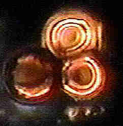
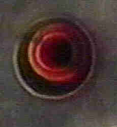
Red Blood Cells Identified in Ground Samples
BROADCAST DISSEMINATION OF TRACE QUANTITIES
OF BIOLOGICALLY ACTIVE CHEMICALS : PATENT
PHARMACEUTICAL COMPOSITIONS
CONTAINING HOLLOW FINE TUBULAR
DRUG DELIVERY SYSTEMS
ADDITIONAL MICROPHOTOGRAPHS OF
BIOLOGICAL AND UNIDENTIFIED COMPONENTS FROM
MAY 7 2000 VIDEO SESSION
Posted June26 2000
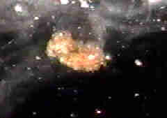
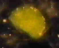
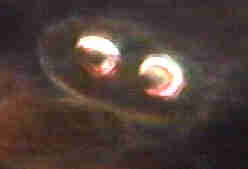

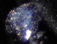
Several of the objects within the video stills from the microscopic session posted within this series remain unidentified. These include the double cells, as well as the blue and green materials shown above. The object in the upper left of this series has been tentatively identified as a white blood cell. Repeated observations of each cell type or object shown here occurred and were recorded within the microscopic video session.
RECENT POST FROM THE SOURCE ON THE MESSAGE BOARD:
MAY 25 2000
Cell Antigen Fixative
I did some digging about the web and found that there are several preparations
that are used to “fix” cells so that thier antigenic structure stays intact. The
antigenic sites or structures on the surface of a cell are the parts of the cell or
micro organism ( infection) our immune system codes to. One particular fixative
is known as Bouins Fix. It’s ingredients are as follows:
2% picric acid – an explosive!
Glacial Acetic Acid
o
37 Formaldehyde
Such chemicals are fairly typical of antigen fixative preparations and are quite
toxic to say the least.
This may account for why some people have reported being burned when
handling some of the material that has been sprayed on us. It would also
account for the sterility of the samples that were recently microscopically
examined. This is a good place to start an analysis if someone so desired.
338glo
RECENT CORRESPONDENCE ON THE MESSAGE BOARD:
MAY 13 2000
Blood Sample Photomicrograph Questioned
Dear Mr. Carnicom:
I read. your post two days ago, and I have some
serious concerns about its validity (not its veracity – I
know that you are an honest man). I believe your
interpretation of the photomicrograph in question is
inaccurate. Your post’s excerpts in “quotes”, my
comments in[brackets].
“Biological components have now been identified in the
two ground samples previously analyzed on
www.carnicom.com.”
[The fact that the subjects evaluated are “ground
samples” sends up a red flag. “Ground samples” could
be samples that are not from aircraft, or, if they are,
could be contaminated by foreign matter. Note that I
am not saying they AREN’T from aircraft or ARE
contaminated, but that they COULD be. In other
words, they were not collected “in situ”, that is,
collected in the air from the contrail itself.]
“An [sic] visual analysis has now been conducted with
a professional quality microscope on May 7 2000….”
[I am not sure what kind of professional microscope
was used, but it appears to me that, based on the
apparent size of the erythrocytes (red blood cells) that
the photomicrograph was taken at between 900X and
1200X. This is borne out by your later statement
“readily visible after being subjected to immersion oil”.]
[However, another inconsistency here is that
immersion oil is not used to provide additional detail to
erythrocytes. Instead, it is used to maintain the same
refractive index between the subject matter and the
objective lens. At the extremely close gap between
objective and subject with a high-power oil immersion
lens, an air gap would cause severe diffraction
problems — thus distorting the view — even with an
fluorite or apochromatic lens.]
[To my experience (and I am not a professional
microscopist), there is nothing that will increase the
‘visibility’ of an erythrocyte. Safranin, methylene blue,
Gram’s, and Gentian Violet stains work wonders, but
only on leukocytes and leukoblasts (white blood cells)
— and I did not see any of them in that picture.]
[But there is a more serious problem. We know that
the microscope used was a optical and not an electron
microscope — else, there would be no mention of
immersion oil (an electron microscope works by
completely different principles). We also know that an
oil immersion lens has a focal length of less than 0.5
mm, which gives a depth-of-focus (or depth-of-field)
in the range of microns. This means that, at the
magnifications involved, you would have almost no
depth perception, and would not be able to maintain
focus past the depth of a single erythrocyte
thickness.]
[Yet we can see in the picture that there are several
layers of erythrocytes, and all are in focus! There is no
way I know of (with an optical microscope) to have
that depth of focus. Also, you can see from the
photomicrographed (especially if enhanced by a
program like Corel Photo-Print or Adobe Photoshop)
that there is no fall-off in the clarity and focus of the
objects.]
[Further, an examination of the photomicrograph, as
well as the shadows in the erythrocyte indentations,
shows that they appear to be top-lighted at an oblique
angle. This is patently impossible with an oil-immersion
lens — it is designed for a light source from either an
Abbe or dark-field substage condenser ONLY.]
[This leads me to believe that the photomicrograph
was made with a methodology that takes advantage of
a technology with which I am not familiar — or it is
faked.]
“The cells appear to be of a freeze-dried or dessicated
nature in their original form within the microscopic
fibers. Isolated and individual blood cells are
interspersed throughout both of the samples that have
previously been described. The surface of the cells
appear to be modified in some way, but electron
microscopy will likely be required to establish further
detail.”
[Based on my limited expertise, I will agree with the
above paragraph to an extent. The erythrocytes
certainly do not exhibit the isotonicity that cells in
fresh blood do, but that’s what happens when blood
dries anyway. I don’t see any evidence for either
freeze-drying or induced dessication, but, like Mr.
Carnicom says, “electron microscopy will likely be
required to establish further detail.”]
“Professional medical analysis of the images and
chemical analysis of the fibers, and the subsequent
disclosure of those results, now exists as a
fundamental need.”
[Absolutely! I have told you in previous
communications that the only thing that will take the
entire chemtrail/contrail business out of the realm of
conspiracy-mongering is for:
(1) Contrail residue to be collected while airborne and
tied to the contrails;
(2) A reputable laboratory to perform the analysis
under strict laboratory methodologies;
(3) The entire analysis, results, and corresponding
data to be published for both lay and peer review.
“The ramifications of this recent discovery establish
sufficient cause for widespread involvement of the
American people in this issue, and for subsequent
Congressional hearings.”
[I disagree. Until we have some serious evidence,
uncontaminated test material, a recognized laboratory
working from valid data, and full disclosure of any
reports (whether they agree with a chemtrail agenda
or not), there is NO sufficient cause for widespread
ANYTHING, and CERTAINLY not for us to spend more
money to send those Bozos in the Congress out on a
tail-chasing exercise.]
[If chemtrails are to be taken seriously, its proponents
must take them seriously themselves, and replace
hysteria, innuendo, gossip, and conspiracy with
evidence, data, and facts.]
“The individual that provided the images herein and
those that will follow shall remain anonymous.”
[That costs him his credibility.]
[Mr. Carnicom, I am one hundred percent convinced
that you are an honest person, but your trust of
people like this will only hurt your own credibility and
give the anti-conspiracy folk more ammunition that
chemtrail believers are all nuts (which I am sure is not
the case). I implore you, for the sake of these people
in this forum if no one else, to provide us with real
evidence if such is available.]
Regards,
Duncan Kunz / duncan.kunz@prodigy.net
A STATEMENT FROM THE SOURCE:
Blood Sample Photomicrograph Questioned
The microscope was a darkfield. The immersion oil was
used directly on the sample to reconstitute the dried
cells. No immersion oil was in a traditional sense except
under the slide to couple the slide to the condensor.
At first water was added to the sample for
observation, but all that showed was the fibers in situ.
The cells in the sample only reconstitute in certain
types of oil. A light machine oil similar to 3 in One oil
was tried with limited success. Also tried was WD 40,
(which was worthless ) and kerosene. The kerosene
did seem to reconstitute the cells, but quickly
bleached them of color. There is a rather sticky
adherent matrix that the fibers are encased in. This
includes the odd blue fiber.
Nearly all the videomicroscopy was done with no more
than a 40X dry objective. A 4X optical coupler is
connected to the video camera. There exists some
portions of the tape that shows individual blood cells
quite clearly. What cannot be seen clearly in the
capture is that this collection of cells is actually
arranged in nice rows. Further, these cells are not fully
reconstituted and are much smaller than their true
size.
There are not as many WBC’s in the sample , but they
certainly do exist. They do not reconstitute well , but
their appearance is unmistakable. Also present are a
least three other types of as yet unidentified cells.
Some with very clearly defined nuclei.
We tried to get a clear picture of the cells in that large
accumulation Clifford posted using the 100X objective,
but were unsuccessful. It was the lack of light passing
through the specimen that limited our ability and not
the actual depth of field. Above 40X, darkfield
objectives must either have a funnel stop or internal
iris. This cuts down on the light dramatically. There are
a few 100X examinations of the material on the video
tape that show individual cells with dramatic clarity.
In spite of the fact that this was collected from the
ground, and in one case off a car that had driven 1000
miles, the sample was sterile! No bacteria , mold or
fungus was found. There was of course some dirt, and
other contaminants in the sample along with some
spores. I do consider the spores to be contaminants
and not part of the original mix.
Two separate samples from different parts of the
United States, dropped on different days, were
examined. Both were identical. One of the samples had
considerably more blood cells, and cells of all types, in
it than the other.
FWIW – there is a method I’ve discovered to enhance
resolution and contrast in any microscope by at least
20 %. It has to do with preparation of the slide itself.
This method was used to prepare all the slides that
were examined.
I hope Clifford posts more captures off the tapes that
he has. Just let me say your jaw will drop when you
see some of what is in that material. My posting this
has more to do with revealing how the cells were
found, than answering your post. It is important that
confirmation can be made by others independent of
me.
[Editors Note: Additional Photographs
Posted May 15 2000;
more to follow]
I won’t get into all the physiologic possiblities that
inhaling blood, or other cells might produce. I’ll just say
for starters that let’s hope this is type “O” blood.
338glo
The following email and subsequent post on the message board was received on June 10
2000:
Hello.
I wish to make a comment on some of the areas discussed, especially
by a certain 338glo. Firstly, fixatives such as Bouins are not used to keep,
“antigenic structures intact.” Fixation causes cross linking of
macromolecules which arrests biological activity, at the same time
rendering the cells amenable to staining. It’s purpose is to
preserve cell ultrastructure to be stained and viewed by light
microscopy, usually by a pathologist, to help diagnos disease.
In other words, 338glo’s implications that your samples might provoke an
immune response is a grossly misleading exaggeration, not to mention
impossible. You will not initiate an immune response to anything that
has been fixed.
By the way:
1)picric acid- is a yellow crystalline substance that is explosive only
when dry and subject to a shock of some kind, such as a blow from a
hammer(not an electric shock). It’s used to precipitate
(separate from solution) proteins, and as a dye.
2)Glacial acetic acid-is just a highly(almost pure) form of acetic acid,
and highly reactive organic acid. %5 is good on your french fries,
99% will burn out all of your mucous membranes. This substance
is used in many laboratory tests.
3)Formaldehyde-another fixative that hardens tissue, and preserves it
for histological examination(staining).
Kunz’s comment about immersion oil is correct. If you want to
reconstitute the cells, try using isotonic saline(approx %0.95 salt in
water). No oil of any kind is going to help you there. You will not
see anything but large structural changes by light microscopy anyway.
You will not be able to judge anything about surface antigens, which, by
the way, were destroyed by the fixation process. That cluster of red
cells looks to be viewed at about x1000. There only appears to be one
fairly visible layer of red cells, if that’s what they are, and not all
of them are in focus. It is impossible to comment on the light source,
but you can view objects under a microscope with light comming from
another source other than the bulb/condenser. As long as it reflects
off the object viewed, travels up the objective to the eye piece, and is
bright enough to see, you will see it. It is also impossible to tell if
the cells(?) were freeze-dessicated or whatever. Cellular specimens can
be frozen, if done correctly, and have their structures preserved; this
statement is nonsense. Also, adding just water(a hypotonic solution)
to those cells won’t help you much either, and will only alter their
structure more if they are at all able to be reconstituted(unlikely).
It is mentioned later that the sample is sterile, BUT it contains
some spores. Do you know what spores are? They’re
a reproductive cell produced by plants and some protozoons. Certain
bacteria(ie: antrax bacilli) also form spores, but
for environmental protection, not reproduction. Another tidbit for
you-type “O” blood accounts for almost half the population,
and is compatible with the other major blood groups, A, B, and AB.
That’s why group “O” is called the universal donor, with
AB being the universal recipient. Inhaling dry blood might make you
cough, but otherwise will do nothing, let alone
initiate an immune response.
This type of fear mongering does nothing but stimulate unfound
paranoia. Please have people that know what they’re talking
about review samples of any significance, or you’ll never be received as
credible. There’s something going on here, and this type of nonsense
isn’t helping anyone find out what it is. I also doubt this sample came
from where you think it did. I hope this helps your readers.
Good luck with the research, and thank-you for
taking the time to read this. I’ve just worked all night, so please
excuse any typing errors. If anything I’ve said needs clarification,
just let me know. Again, thanks.
Shayne Dixie M.L.T.
(Medical Laboratory Technologist)
Brockville General Hospital
Brockville, Ontario Canada.
K6V 1S8
(in case you wonder where I’m coming from)
A STATEMENT FROM THE SOURCE:
Posted June 14 2000
On June 12, Shayner made a post and my first reply
was lost when I tried to post it. Hopefully this
individual message will post.
Bouins fix has many uses, please see the following
quotes from the listed web sites for it use as an agent
to fix antigen sites on cells.
www.emsdiasum.com/ems/his…ative.html
Bouin’s Solution
Bouin’s solution can be used as a fixation and a
staining fluid. Bouin’s
fixative is excellent for use on biopsy specimens of the
gastrointestinal
tract. Tissue from the endocrine system are well fixed
and many antibodies
react well with tissue fixed in Bouin’s.
www.cmbm.org/Conference98…s/205.html
Next one, please. We can detect the cancer cells by
immunocytochemistry
using the same type of antibodies. Here we have tumor
cells detected with
one of the antibodies. This was again the free beta
subunit. The way in
which this tissue is fixed for cytochemistry is different.
We don’t fix in
formalin. To have a result in immunocytochemistry,
when you’re dealing with
hCG, aldehydes kill glycolipids and carbohydrates, so
we have to use a
picric acid type of fixative, Bouin’s fixative
www.alzforum.org/members/…table.html
Immune system response occurs for many reasons, and
involves many different methods. The deciding factor
in initiating an immune response is that something
foreign has come into contact with the interior of the
body, or the external mucous membranes of the body.
This could be a piece of wood, dirt, metal, mold,
bacteria, viruses, pollen, chemicals, or cellular tissue
for example.
Many people have seasonal allergic responses to
pollens, grasses, molds, and so on. Some people have
inhallation type allergies that will initiate asthmatic
bronchospasms that can be life threatening. Some of
the recent postings regarding eye, throat, and lung
irritation following spraying are typical of an immune
system response. Foreign blood cells of the wrong type
will cause an immune system response. If the outer
membrane of the cell has been changed in some
manner this will cause an immune system response.
Foreign cellular tissue will cause a dramatic immune
system response. Transfuse the wrong blood type,
implant a new organ, and your immune system will
immediately go to work, clot the blood,and reject the
organ.
I mentioned Bouin’s fix as merely an example of a
known fixative agent that will preserve antigenic sites
on blood cells. The ingredients in the mixture are all
toxic, some are known carcinogens and one when dried
is explosive. This is a place to start analysis of the
material. Samples of this stringy , sticky material have
been collected by people across the US. There are
reports of physical contact causing people to become
ill and thier skin to be burned. Some of ingredients in
Bouin’s fix could easily cause such symptoms.
Here is the EPA haz mat paper on Bouin’s fix.
www.mastertechs.com/msds/…FXBOU2.TXT
C. FIRST AID (EMERGENCY PROCEDURES)
1. EYE CONTACT: Remove contact lenses if necessary,
and immediately flush
eyes with copious amounts of water. Buffered saline
eye wash
solution may also be used. Seek medical attention
immediately.
2. SKIN CONTACT: Immediately wash contaminated
area thoroughly with
mild soap and water. Remove contaminated clothing if
necessary.
3. INHALATION: Remove to fresh air. If symptoms
persist, seek medical
attention.
At the moment of course there is no proof that the
cells in the sample have been treated with any fixative
agent. Nor is there any proof that any of the
ingredients in Bouins fix are in the sample. This needs
to be determined. Mr. Carnicom spent over 400 dollars
of his own money having a government licensed lab
analyze some of the samples. The report he recieved
was essentially worthless. [Editors Note: Lab fees of
$450 were paid for the lab tests referenced, however,
I was not personally the source of those funds. CEC]
The reason immersion oil was used, is that the sticky
matrix that held the cells would not dissolve in water.
Water was useless to dissolve the matrix and allow the
cells to reconstitute. Immersion oil however worked
very well for this purpose.
My comments about the spores were only that due to
thier paucity, I felt that these were contaminents and
not native to the sample.
My posting was not to incite fear, it was to promote a
starting point to analyze what has been sprayed on
some parts of the US. The facts are that this material
has been seen and collected from different sites
around the US, and like it or not it does exist.
338glo

