
BLOOD ISSUES INTENSIFY
Clifford E Carnicom
Apr 22 2009
I am not offering any medical advice or diagnosis with the presentation of this information. I am acting solely as an independent researcher providing the results of extended observation and analysis of unusual biological conditions that are evident.
Three independent methods have been established that appear to confirm the presence of developing modified erythrocytes (red blood cells) within cultured dental samples that exhibit the characteristics of the Morgellons condition as previously researched and identified. All individuals tested thus far have produced the dental filamentous materials, regardless of whether visible skin anomalies are present or not. Please see previous research for further clarification on the prevalence of the condition.
The erythrocytic detection methods are:
1. Direct observation under the microscope at relatively high magnification (8000x – 10000x) using developed microscopy techniques.
2. The use of the Kastle-Meyer presumptive test (visual and microscopic, sensitive test) for blood, a method commonly used in forensics for blood identification.
3. The HEMASTIX (TMP) presumptive forensic test (very high sensitivity) commonly used for blood identification.
The tests have been repeated several times to assure consistency in methods, results and controls.
The appearance of the cultured erythrocytic cellular structures, if accepted as properly identified, in and of itself defies all conventional understanding of blood cell development. This appearance also corroborates a long history of research through this site of environmental and biological samples that defy conventional expectations and knowledge with respect to the state of public health and the environment (e.g, refer to Extraordinary Biological Observations, Carnicom, May 2004). Simply put, erythrocytes are not to be grown in the the test tube under the current state of conventional knowledge. To do so, however, is considered to be a holy grail of biological achievement with huge implications for bioengineering, human health and the human species. Ground breaking research in this aspect of biology, i.e, the “growing of blood cells” has been reported in the media throughout this last year, and was simultaneously stated to entice immediate interest from the Defense Department for battlefield applications (radio news report). Research previous to this recent announcement reports the sustenance and perpetuation of existing cells within a growth medium, but not the creation of new cells. Achievements of growth on any scale are clearly on the leading front of stem cell research, and comparative questions must be raised regarding the state of public disclosure on the subject vs. actual technological achievements that may already be in place.
I have no desire to sensationalize this subject as the seriousness of the issue is apparent to those that understand the ramifications of this report, should it bear itself to be true. I am obligated, however, to report on the state of affairs as they are encountered through honest research. I would hope that all three methods used here along with all previous reports involving erythrocytes for more than 10 years can be shown to be false, but if so, it will have to be done with open and public research that is subject to full cross-examination. I would prefer to not be forced to continue to report findings of this nature but the obligations with respect to public health and the environment do not afford me that liberty.
It has taken some time and effort for me to be able to employ three independent methods of erythrocyte identification at the forensic level, but the seriousness of the subject requires this as a minimum. I do not state this subject to be a closed affair; to the contrary, I am opening a door that requests that there be additional resources activated to conduct the investigations in proper earnest. The purpose of this paper is not to incite controversy. It is to acknowledge what appears over and over to be a very real issue that appears to be of consequence whether we would like to confront it or not.
From the vantage point of this researcher (through varied research over an extended period), it is difficult to come to any other interpretation than that the Morgellons condition is very likely to be fundamentally a blood borne condition. It is quite possible that the findings of this report demonstrate a key element of the Morgellons condition. From additional extensive research that has been conducted, it appears likely that it affects the general population at large. Any skin anomalies or surface manifestations appear to be just that, and they are not necessarily representative of the underlying causative factors. It also appears unreasonable to use surface or skin manifestation as a primary criteria for assessing the extent and distribution of the condition. It may be wise to consider the blood condition of the general population as a focal point of further investigation and research. The associations of airborne and environmental factors also established through extensive research must also be given their due consideration.
ADDITIONAL DETAILS:
Culture Development:
|
|
|
|
||
|
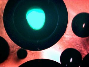
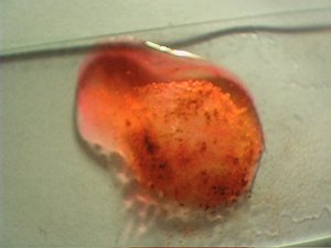
Original extracted filamentous dental sample in red wine contained and held in a petri dish with lid. This acts as the culture medium of this report. This sample represents a total collection period of nine minutes of gum exposure to the wine solution; three segments of three minutes each, respectively. |
After approximately two to four weeks, a filamentous growth form emerges on the surface of the wine (unanimous at this point). This growth form is identical in nature at the microscopic level to previous dental cultures that have been reported on with the use of an agar medium. A time lapse video of that growth is available on a previous report. |
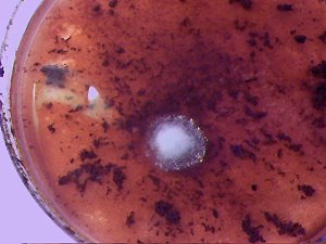
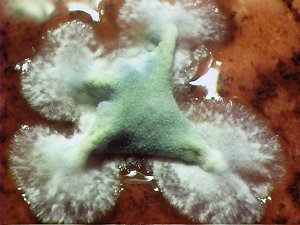
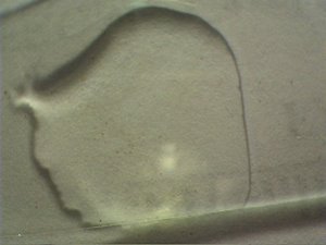
The earlier forms of the filamentous growth are pure white as in this sample. This is identical to previous agar medium cultures that have been developed. At this stage, the growth is approximately two to three days old. The photographs in this collection are taken from more than one culture to demonstrate various stages of growth and development. |
||
|
|
|
|
||
|
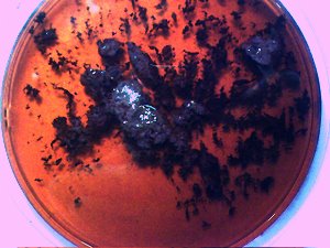
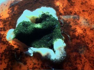
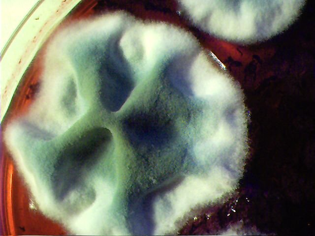
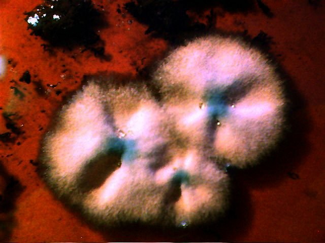
The culture growth several days into development. This stage produces greater variation in the surface structure. Additional structure and color is introduced and appears visually to be more of a mold or fungus nature. It is this stage which departs from the previous agar cultures and adds greater complexity to the form. It appears that the nutrients within the wine are essential to reaching this stage of development,. |
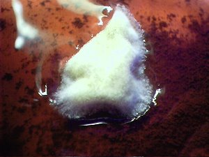
Further development of the filamentous form. A convoluted surface is now a characteristic feature. Some structures have been observed to reach approximately one inch in thickness with numerous folds before being constrained by the lid of the petri dish An additional difference between the agar medium and the wine medium is that growth on the agar medium can occur within 24 hours, whereas the wine medium requires approximately two to four weeks to begin the growth process.. |
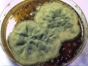
The final form of development that has been reached by the culture growth. Total time elapsed at this stage is approximately one to 1 1/2 months after the appearance of the original growth. Total time elapsed is therefore on the order of two to three months after the original collection of the dental sample in the wine medium. The growth form reaches a much greater degree of complexity than with the use of the agar medium. The diameter of the growth is approximately three inches and appears to be constrained only by the petri dish boundary and available nutrients. The structure is cohesive. Thus far every dental sample observed has produced this growth form and only this growth form after the time lapse of two to three months. |
Microscopic Analysis:
|
|
|
|
||
|
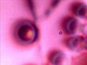
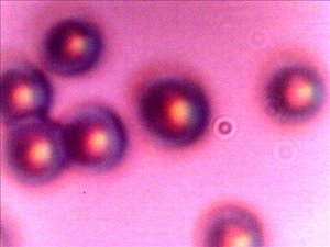
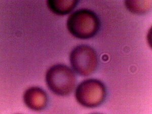
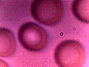
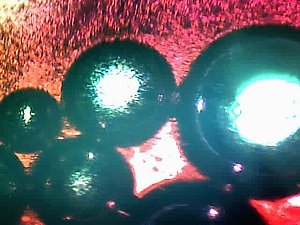
What appears as a fully reconstituted ethrocyte on the left side of the photograph. Diameter approximately 6 microns. Biconcavity of structure is apparent. A reconstitution process similar to that reported earlier on eyrthrocytic studies appears to also be in place within the wine medium. Partial reconstitution appears to take place in the wine medium, additional reconstitution appears to take place over a 20 to 40 minute period under the light and heat of the microscope stage. Additional partially reconstituted structures on the right side of the microphotograph. Magnification approx. 9000x. |
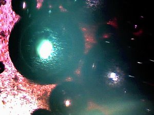
A set of largely or fully reconstituted erythrocytic structures. Biconcavity characteristic of erythrocytes is apparent. Original size at time of first observation is on the order of 4 to 5 microns in diameter. Full biconcavity and uniform shape not apparent at beginning of observation period; this develops more fully under on the microscope stage as sample is subjected to additional light and heat. Reconsitution takes place to a size range of 5-6 microns with largely uniform structure under these conditions. Human blood cells are on the order of 6-8 microns in diameter. Additional red blood cell size comparisons are available at Biological Observations Confirmed, Carnicom, 2001. Magnification approx. 9000x. |
A set of partial and mostly fully reconstituted erythrocytic structures.The non-reconstituted form is more typical of early observation in the session; it appears as though additional heat and/or light is responsible for the final reconstitution that takes place. No budding or fission process characteristic of yeast cell reproduction is observed. Biconcavity and size range is a unique identifying characteristic of erythrocytes. If erythrocyte identification is accepted, species of blood remains unidentified. It can also not be be stated that the reconstitution process is entirely complete. Magnification approx. 9000x. |
||
|
|
|
|
||
|
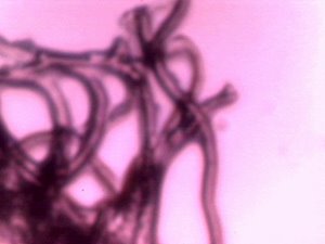
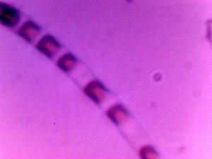
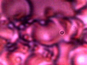
Two main structural forms are visible with the culture growth: filaments and erythrocytic forms. This microphotograph presents the filament aspect of the sample. Dimorphic fungal forms such as Candida have also been a serious topic of consideration in this study. Reconstitution, biconcavity and lack of budding or fission observed fails to support the conventional fungal hypothesis. Additional forensic studies were conducted to eliminate ambiguity and they further confirm the tenets of this report. Additional similar findings under different circumstances over the history of research on this site must now be be given further consideration. Magnification approx. 2000x |
A combination of a filament section with surrounding erythroctyic structures. There does appear to be a relationship between the presence and origin of the presumed erythrocytes and the filament forms. This relationship is not clearly defined at this time. There are some indications that the filaments may be the source of origin for the presumed erythrocytes, but actual formation has not been observed.. It can be stated that there is no fission or budding that has been observed to date. Time lapse imagery may shed further light on this issue. Magnification approx. 1500-2000x. |
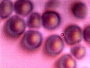
A good example of variation in the reconstitution process. Three primary changes take place during the reconstitution stage: First, the size of the presumed erythrocye increases, as if a dessication stage may have been breached. The second is that uniformity of circular form develops. Lastly, the biconcavity characteristic of erythrocytes develops. This image shows examples of all three of these stages. The structure at the lower left is in the original form observed. The structure near the top and the structure to the lower right have increased in size, but they do not yet exhibit the biconcave stage of development. The central two structures show reconstitution with increased size and biconcavity visible. It is appropriate to compare the two central structures with the control image of a human blood cell in this series below. Magnification approx. 9000x. |
||
|
|
|
|
||
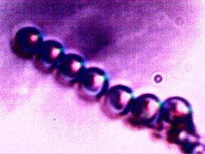
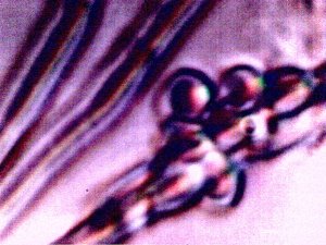
| A peculiar, but not unique example of structures within the filament form. It is this image which brings to question the origin of the presumed erythrocytes. It is also of interest to compare this image with the one that immediately follows to the right. Magnification approx. 9000x. | One of the early observations which promoted further detailed study. The co-linear aspect of development raises further questions about the association and relationship between the filament form and the erythrocytic form. It is not expected that erythrocytes in any natural arrangement would display this type of alignment. At the lower right of the photograph may be evidence of a filament or vestiges of a filament. It was during this observations that reconstitution was also observed, as biconcavity increased during the observation session. Magnification approx. 9000x. | A representative example of the combined filament and erythrocytic forms. Magnification approx. 9000x. | ||
|
|
|
|
||
|
A control example and microphotograph of fairly uniform human blood cells, or erthrocytes. The only visual difference between this control photograph and the reconstituted structures that are the basis of this report is that of size. Human erythrocytes generally measure on the order of 6-8 microns in diameter. The erythrocytic structures of this report, after apparent full reconstitution, are on the order of 5-6 microns. A human hair is on the order of approx. 60-100 microns in thickness. The presumptive forensic tests of this report do not establish a case of human blood, only that of blood and hemoglobin. Questions of what is full reconstitution under optimal environmental conditions and consideration of species involved remain open questions. Magnification approx. 9000x. |
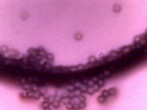
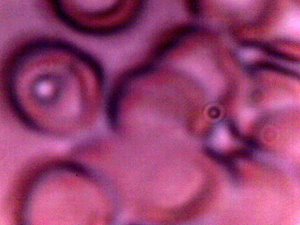
An example of the anomalous blood condition that has been the subject of numerous reports on this site. The individual providing this blood sample produces a relatively large amount of the dental filaments during the wine extraction process. The culture that developed from this individual was the quickest to appear; approximately two weeks were required vs. what can be up to four weeks for the other cases. The culture growth from this individual was also extensive and rapid relative to other individuals. The individual displays no known skin anomalies. At this point, there appears to be a fairly strong and direct correlation between the conditions of the blood that have been observed along with anomalous physical manifestation, whether it be skin anomalies or the volume of dental materials removed. Please see additional reports on this site for further description of the blood condition that is depicted here. Magnification approx. 9000x. |
Another example of the anomalous blood condition that has been described on numerous reports on this site. Deformation of the cellular structure is apparent and common. This individual is the same as that of the previous photograph. Bacteria is also a strong consideration in this case; please refer to previous references to Chlamydia Pneumonia and its characteristics. Essentially all individuals that have been observed in these studies show degree of these anomalies, regardless as to whether skin anomalies are present or not. All individuals tested thus far produce some degree of the dental filaments. All cultures mediums established thus far produce the culture form that is the subject of this report. Magnification approx. 9000x. |
||
All magnifications reported are prior to reduction of images for web site presentation.
Kastle-Meyer Blood Detection Forensic Test:
( Performed on Microscope Slide)
|
|
|
|
|
View of cultured dental sample #1subjected to Kastle-Meyer blood detection forensic test. Peroxide activity characteristic of hemolysis and phenolphthalein color change to bright pink-red is evident. A positive presumptive test for the existence of blood within the cultured dental sample. On glass microscope slide.The sample is dried prior to performing the test. Magnification approx. 2x. |
View of cultured dental sample #2 subjected to Kastle-Meyer blood detection forensic test. Peroxide activity characteristic of hemolysis and phenolphthalein color change to bright pink-red is evident. A positive presumptive test for the existence of blood within the cultured dental sample. On glass microscope slide. The sample is dried prior to performing the test. |
|
|
|
|
|
|
Control case for the Kastle-Meyer presumptive blood detection test. View of human blood sample subjected to Kastle-Myer blood detection forensic test. Peroxide activity characteristic of hemolysis and phenolphthalein color change to bright pink-red is evident. A positive test result. On glass microscope slide. The blood is dried prior to performing the test. Magnification approx. 2x. |
Second control case for the Kastle-Meyer presumptive blood detection test. No blood present in the sample. No peroxide hemolysis evident and no color change. A negative Kastle-Meyer forensic test result. On glass microscope slide. Magnification approx. 2x. |

Summary : Both the human blood cell control test and the cultured dental samples produce the same visible physical
and chemical reactions at the visible level and satisfy the expected conditions of blood detection by the Kastle-Meyer forensic test.
Kastle-Meyer Blood Detection Forensic Test:
(Microscopic View)
|
|
|
|
||
|
Microscopic view of human blood sample subjected to Kastle-Meyer blood detection forensic test. Peroxide activity characteristic of hemolysis and phenolphthalein color change to bright pink-red is evident. |
Microscopic view of cultured dental sample #1 subjected to Kastle-Meyer blood detection forensic test. Peroxide activity characteristic of hemolysis and phenolphthalein color change to bright pink-red is evident. |
Microscopic view of cultured dental sample #2 subjected to Kastle-Meyer blood detection forensic test. Peroxide activity characteristic of hemolysis and phenolphthalein color change to bright pink-red is evident. |
Summary : Both the human blood cell control test and the cultured dental samples produce the same visible physical
and chemical reactions at the microscopic level and satisfy the expected conditions of blood detection by the Kastle-Meyer forensic test.
HEMASTIX (TMB) Blood Detection Forensic Test:
|
|
|
|
||
|
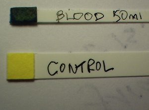
HEMASTIX control blood test. The lower stick is that of a negative, i.e., no blood of any trace evident. The upper stick is a positive test that corresponds to one to two drops of human blood in 50 milliliters of distilled water. The HEMASTIX test is positive for the existence of blood essentially if any green to blue-green tint shows on the stick after a time interval of 60 seconds. The HEMASTIX test (TMB) is highly sensitive to the existence of blood in a sample. Magnification approx.2x. |
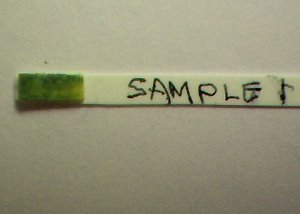
Positive HEMASTIX test result when |
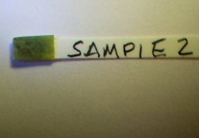
Positive HEMASTIX test result when |
Summary : Both the human blood cell control test and the cultured dental samples produce the same visible physical
and chemical reactions and satisfy the expected conditions of blood detection by the HEMASTIX forensic test.
FIRST GROWTH INHIBITION STUDY:
An initial growth inhibition study follows below.
This information is of a preliminary nature only and the details of the study will not be presented at this time.
Copper sulfate can be toxic or lethal to the human species in sufficient quantity.
I repeat that no medical advice or diagnosis is presented in this paper.
NO ONE is advised to experiment with the ingestion of copper sulfate under any conditions as a basis of this report.
All individuals are advised to consult with a medical professional for any medical related issues.
This presentation is for information purposes only.
|
|
|
|
|
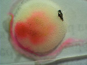
Control Culture. Growth is rapid, extensive and described as above. |
A separate culture exposed to a solution of copper sulfate and the original wine base for approximately three days. Growth of the culture appears to have ceased at the time of exposure to the copper sulphate. Over the next several days, discoloration of the culture takes place to a red-brown cast. Additional study is required. Details of the study will follow later as time and circumstances permit. |
Additional Note:
The Carnicom Institute now exists as a non-profit corporation registered in the state of New Mexico.
Those that wish to support the mission and goals of the Carnicom Institute may make contact at the following web address:
http://www.carnicominstitute.org
or at:
Carnicom Institute
PO Box 355
Wallace, ID
83873
USA

