
MORGELLONS:
PATHOGENS & THE GENERAL POPULATION
Clifford E Carnicom
Mar 08 2008
Last Edit Apr 09 2008
I have no medical expertise and I claim none. I am not offering any medical advice or diagnosis with the presentation of this information. I am acting solely as an independent researcher providing the results of extended observation and analysis of unusual biological conditions that are evident.
There is increasing evidence that the general population may be affected by at least four pathogenic forms. Any perception that only a small segment of the population is affected by the so-called Morgellons condition may be quite false. Certain pathogenic forms are repeatedly showing up in a majority of human biological samples that have been observed, regardless of whether or not visible skin anomalies appear. The use of unusual skin conditions as a defining criteria of the existence of the “Morgellons condition” appears to be completely inadequate and it does not appear to encompass the severity, extent and distribution of the pathogenic forms.
The pathogenic forms are as follows:
1. An encasing or encapsulating filament, often barely visible to the human eye. This filament form often measures on the order of 12 to 20 microns in thickness. This bounding filament form usually contains within it a network of sub-micron fibers that are generally in parallel alignment with one another. This filament form has been found within airborne, skin, saliva, gum(dental) and ear wax samples. It has also been observed within an anomalous hair sample. It appears possible that the function of this particular form is to deliver or encase a sub-network of additional pathogens of much smaller size. No natural identity of this form has been established.
2. A network of sub-micron filament forms. These are usually encased within the bounding filament referred to above. A human hair is on the order of 60-100 microns in diameter; an asbestos fiber is on the order of 2 microns in thickness. This pathogenic form has repeatedly been found within airborne, skin, saliva, ear wax and gum(dental samples). This form has some morphological similarity to fungal forms (i.e., hyphae) but no suitable match to any known species exists at this time. In addition, there is no match at this time to a eukaryotic cell structure which is required for a match to fungus. A “budding”, or apparent growth structure composed of filaments of this same class has been identified on skin borne biological samples. A method for the testing of chitin, usually present in the cell walls of fungi, is to be established.1
3. A sub-micron spherical to oblate structure. The best size estimate for this form is currently on the order of 0.5 – 0.7 microns. These structures can and often do occur in large numbers within the biological samples. They have been found in isolation within the bounding filament as well as in large concentrations within the bounding filament. The have been found in combination with the sub-micron fibrous network within the bounding filament. They have been found in airborne, skin, saliva, ear wax, gum(dental) and anomalous hair samples. They have been found within a broad distribution of human blood samples. They can and often do exist as an intracellular(within the cell) organism. The damage to cellular integrity(i.e, erythrocytes, or red blood cells) appears to correspond directly to the numbers present of the organism. The best assessment to date that can be offered by this researcher is that of a Chlamydia-like(coccus) bacterial form, with special a emphasis upon Chlamydia pneumoniae. The modification of conventional biological forms or of pathogens to exotic levels should be strongly considered throughout this investigation, in addition to the creation of new or unknown forms. The structure stains Gram-negative.
4. What is being called, for the time being, a “hybrid” form. This form has properties that are somewhat in between the sub-micron Chlamydia-like form and the sub-micron filament form. This form appears to be a state of transition between the two more defined sub-micron forms. It has been found only in biological samples and not in airborne samples. It occurs commonly within, but it is not restricted to, human blood samples and it often is in association with the Chlamydia-like form. It is of generally filamentous form but it is reduced in length compared to the sub-micron fibrous network that has been itemized. Mycoplasma or Mycoplasma variations or modifications are one consideration within this topic.
Readers will find numerous microphotographs of the mentioned forms on a series of papers that have been recenly presented on this site, as well as in the examples presented below. These pathogenic forms were first and originally identified within two subjects that manifest visible “Morgellons” skin symptoms, and it may have been surmised that such pathogens are restricted to that class of individuals.
The remainder of this paper will present evidence that visible skin anomalies are not a suitable criteria to establish the existence of the so-called “Morgellons condition” and that certain pathogenic forms may be repeating internally within a broad cross-section of the general population. The results from four individuals will be presented. These individuals were selected essentially at random and they possess NO VISIBLE SYMPTOMS that are commonly being reported as characteristic of the “Morgellons condition”. Every individual that was tested in the manner of this report(gum-dental & blood) demonstrates the existence of these pathogens within their body. The prevalence of the four [SEE NOTE BELOW] pathogenic forms in all cases thus far appears to be indiscriminate as to age, sex, or the general state of visible health for that matter. It appears that the “Morgellons condition” has been defined primarily in terms of anomalous and visible skin conditions; it is apparent that this restriction is artificial and essentially meaningless with respect to the existence of the pathogenic forms described here. The only correlation that may be made with visible symptoms appears to relate to the number or extent of pathogens that are present in the body.
Put more bluntly, the general public may wish to become engaged in this issue.
The following is a set of microphotographs from individuals that do not display any visible symptoms (i.e,, outwardly visible) of the so-called “Morgellons condition.” These photographs are at high magnification and they are at the limit of visible microscopy.
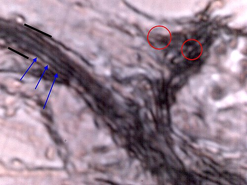
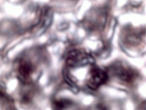
Subject No. 1. Male subject aged 55 years. No visible “Morgellons”symptoms.
Dental-gum expelled sample. Sample extracted with use of hydrogen peroxide-red wine mix.
(please refer to Morgellons: A Natural Medicine Approach, by Gwen Scott N.D.)
Three pathogenic forms visible: bounding filament (black border),
sub-micron interior filament network (blue arrows) and Chlamydia-like organisms(red circles).
These same pathogenic forms repeatedly found in an individual with
visible skin anomalies characteristic of the so-called “Morgellons” condition.
Magnification approx. 7000x.
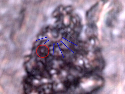
Subject No. 1. Male subject aged 55 years. No visible “Morgellons”symptoms.
Dental-gum expelled sample. Sample extracted with use of hydrogen peroxide-red wine mix.
Chlamydia-like organisms visible(red circle) and “hybrid” form(blue arrows).
The Chlamydia-like organisms measure at approx. 0.5 microns.
Magnification approx. 7000x.
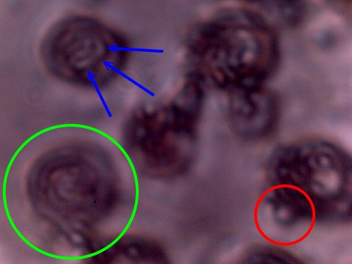
Subject No. 1. Male subject aged 55 years. No visible “Morgellons”symptoms.
Blood sample that has been subjected to the Gram stain process to identify bacterial forms.
Erythrocyte (red blood cell)(green circle) cell structure is expected to and will deteriorate as a result of the stain process.
Unstained erythrocytes measure on the order of 6-8 microns.
Chlamydia or Chlamydia-like organisms(red circle) are evident and more readily visible as a result of this stain process.
They can, with careful observation, be observed to disrupt the topography or surface of unstained blood cells;
this is a further indication of the intracellular property of the organism.
Chlamydia-like organisms measure on the order of approx. 0.5 – 0.7 microns.
Hybrid form also visible in this stained sample(blue arrows).
Organisms are intracellular, i.e, they exist within the cells.
Bacterial infections within the blood can be of grave consequence.
Magnification approx. 7000x.
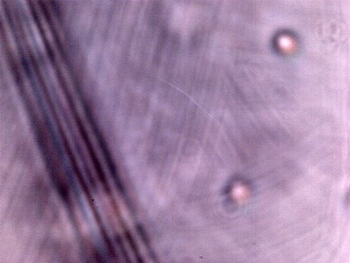
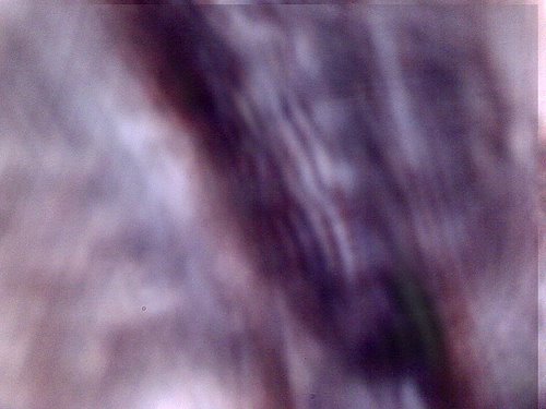
Subject No. 2
Male, approx. 60 years.
Dental sample. Encasing filament and sub-micron fibrous network.
Magnification approx. 7000x.
Subject No. 2
Blood cells subjected to the Gram stain process. Blood cell structure is damaged as a part of the stain process
and the Chlamydia-like structures become visible as a result.
Magnification approx. 7000x.
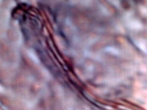
Subject No. 3
Female, 60 years.
Dental sample. Encasing filament and sub-micron fibrous network.
Magnification approx. 7000x
.
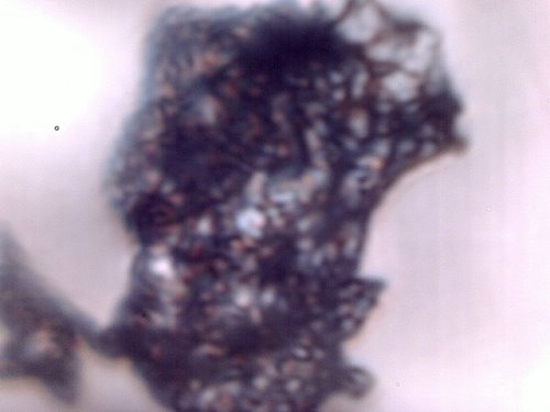
Subject No. 3
Female, 60 years.
Dental sample. Extensive collection of sub-micron Chlamyida-like structures..
Magnification approx. 7000x.
Subject No. 4
Female, approx. 60 years.
Dental sample. Sub-micron fibrous network.
Magnification approx. 7000x.
Additional Notes:
Twenty-six, not four, individuals as a minimum have now subjected themselves to the sampling method of this report. The sampling process now crosses numerous state lines, including New Mexico, Colorado, North Carolina and Montana. Twenty-six of the twenty-six subjects show the same pathogenic forms. The above photographs document only four of the fourteen individual samples. Most of the individuals show no outwardly visible forms of affliction. The only variance is in the amount of material that exists or is produced within the body. Visible skin signs of the “Morgellons” condition are not a suitable criteria to establish the existence of the pathogenic forms in the body. The evidence supports the contention that the general health of the population has been seriously compromised by the pathogenic forms under examination.
My time available for research and the presentation of the information remains limited. This website exists on a month-to-month contract. The existence of this website is not guaranteed. There is no known independent off site copy of this website. The public should preserve, protect and distribute the information on this site to their own level of confidence and assurance.
Clifford E Carnicom
Feb 13 2008
Edit Apr 09 2008
References:
1. Chemical Analysis for Chitin as a Measure of Fungal Infiltration of Cellulosic Materials., Army Mobility Equipment Research and Development Command, Fort Belvoir, Defense Technical Information Center, Access No. ADA036986, http://www.dtic.mil/dtic/tr/fulltext/u2/a036986.pdf

