
Environmental Filament Project :
An Introduction
Clifford E Carnicom
Jul 09 2013
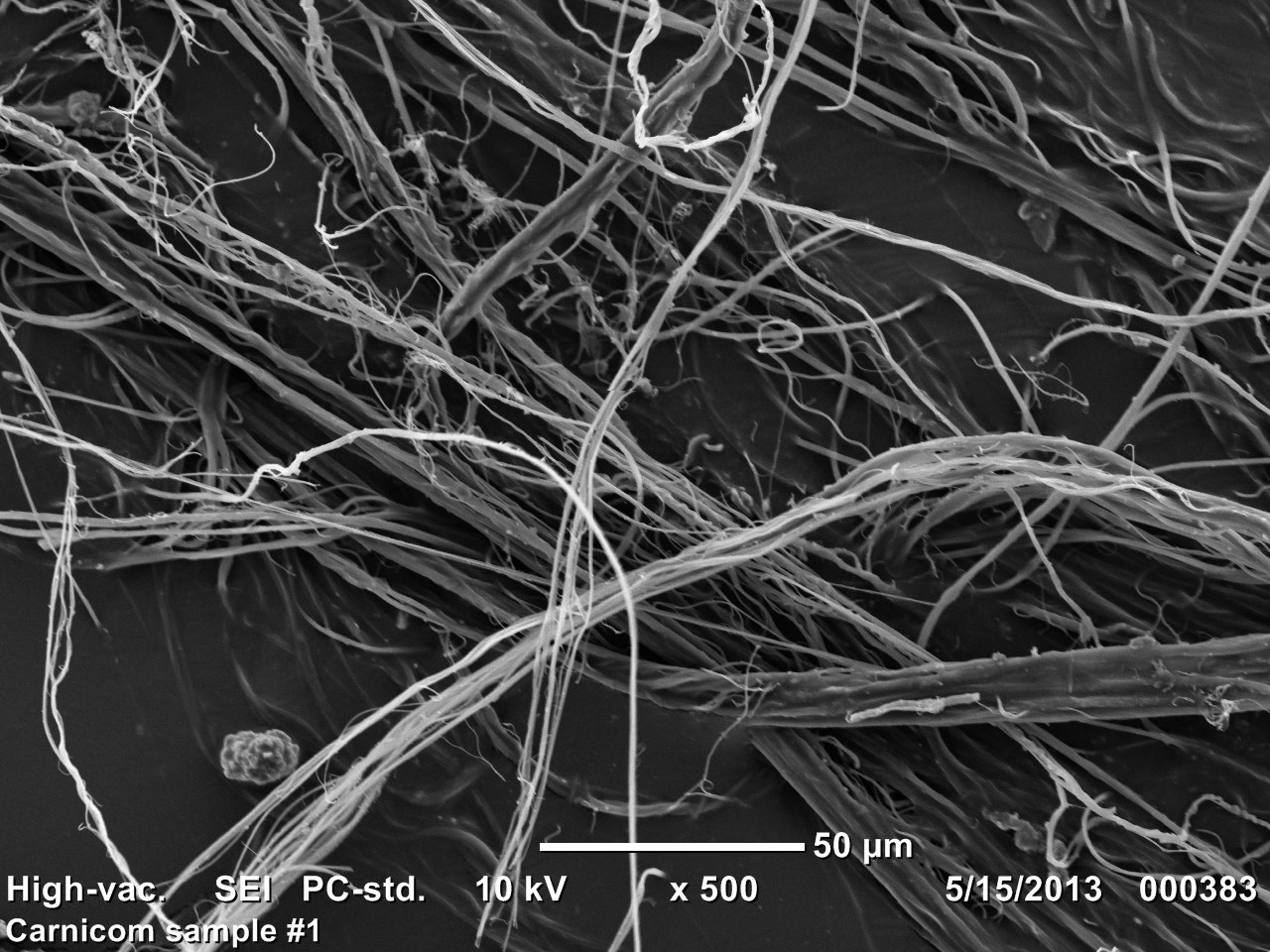
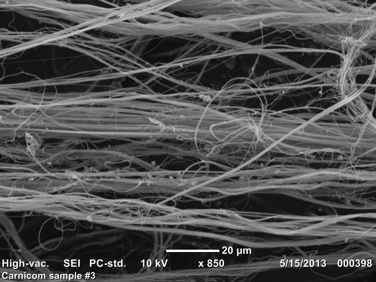
Under current projections, it wll be some months ahead before I will be able to engage fully into the Environmental Filament Project that has been outlined under this site. In the interim, however, an important introduction to what lies ahead can be presented. Carnicom Institute is now able to display a series of scanning electron microphotographs of a typical sample; they will not be discussed in any detail until I am able to begin the study project. Those familiar with my work may be aware of my reluctance to use the term nano-technology in association with any environmental or biological samples examined thus far; this has been due to the lack of any electron microscope images that are derived directly from these same samples. This is no longer the case, and the use of the nano-technology term in association with this material is now fully justified. The samples shown below are identical to those that the United States Environmental Protection Agency has refused to identify or analyze. It has taken close to a decade and a half to acquire these images; appreciation is extended to all parties that have helped to make this information available to the public. Sufficient additional samples have been received, both national and internationally, to support the Institute project plans. This study will begin as the opportunity affords itself and as parallel work that is underway is completed. Light microscope images of the same material are also shown below.
Carnicom Institute : Electron Microphotographs of Environmental Filament Sample


Carnicom Institute : Light Microscope (CMOS) Photographs of Environmental Filament Sample
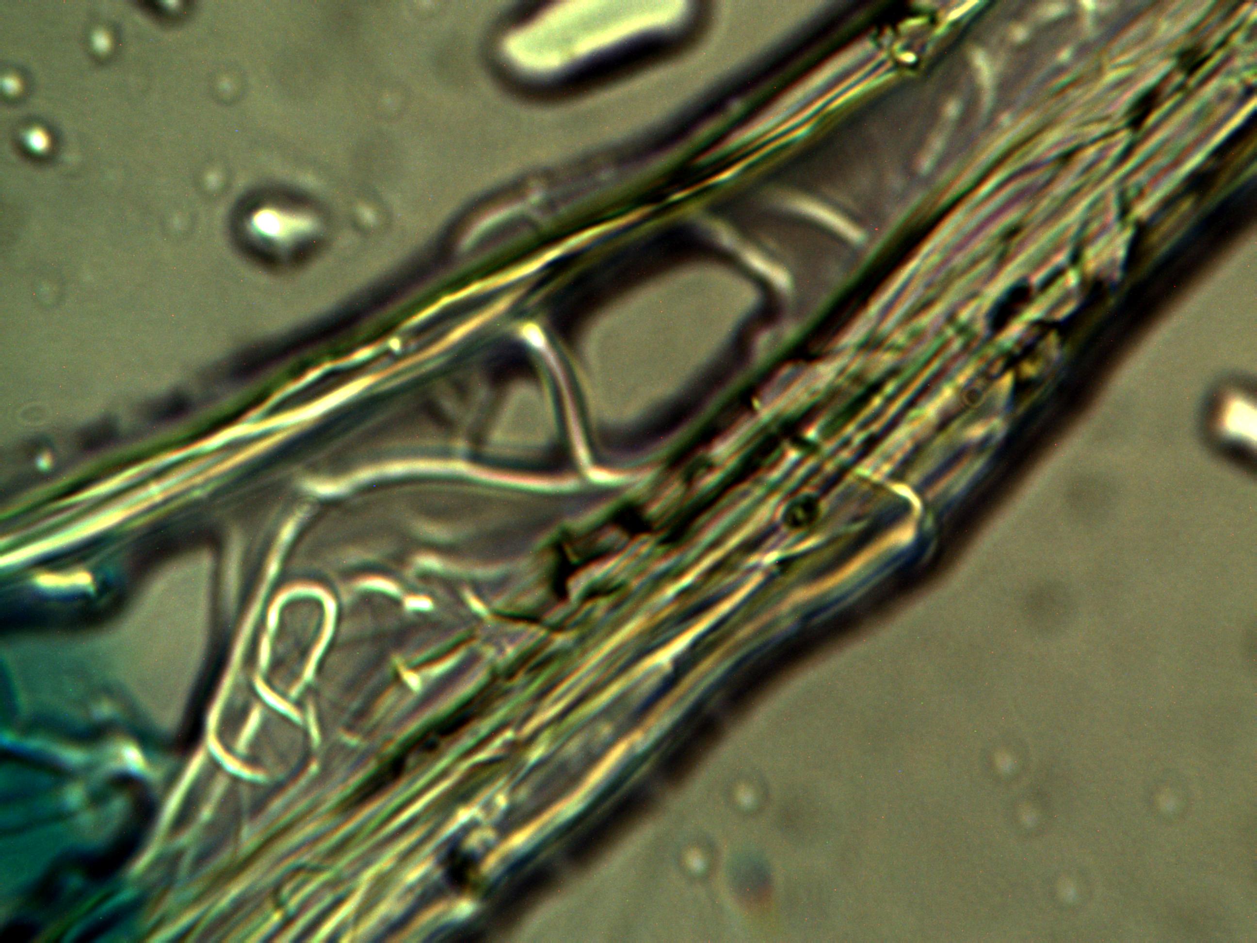
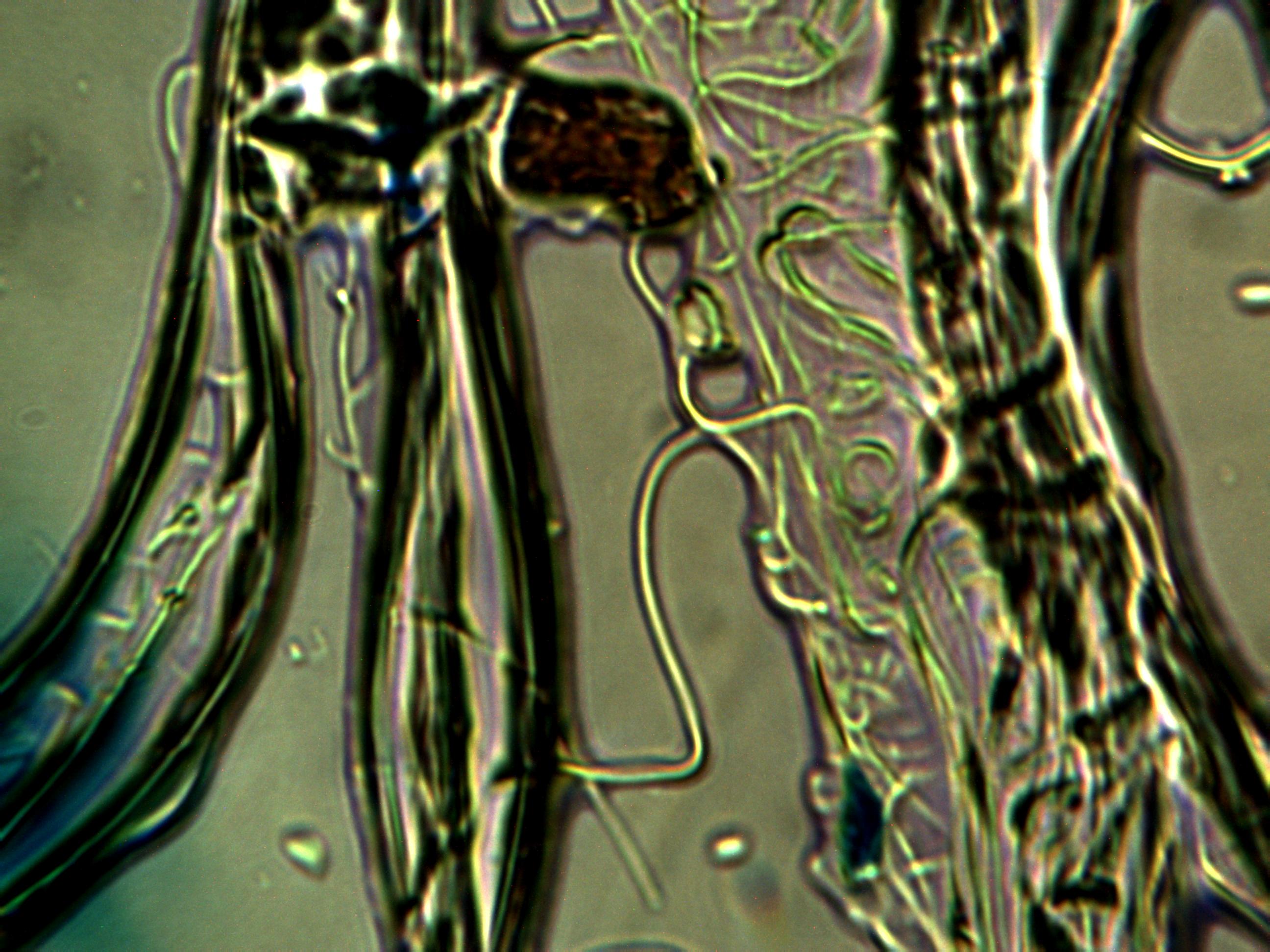
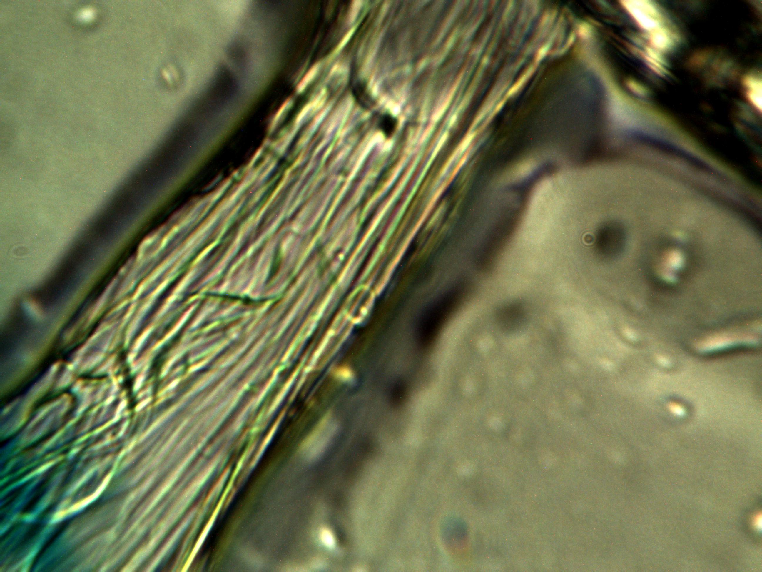
Approximate magnification of original imagery : 6000x
