
ARTIFICIAL BLOOD (?)
Clifford E Carnicom
Aug 27 2009
I am not offering any medical advice or diagnosis with the presentation of this information. I am acting solely as an independent researcher providing the results of extended observation and analysis of unusual biological conditions that are evident.
Strong evidence now exists that an artificial or modified blood form is a dominant internal component, if not the dominant component, of dental filament samples that are commonly associated with the Morgellons condition.
A method has been developed that breaks down the external casing of the fibers. A reconstitution process then takes place. The constituents in the resulting solution have been repeatedly examined under the microscope at high power. The method has been replicated numerous times, and on each occasion the same identifiable structures result. The structures indicate that they are a form of erythrocyte, or red blood cell.
It has been repeatedly proposed by this researcher that the condition of the blood appears to be a common denominator of the Morgellons condition; this latest research further substantiates that position. Essentially all individuals tested thus far demonstrate these same blood variations to some degree, regardless of whether certain skin anomalies are present or not.
It has previously been established that cultures developed from the dental samples are also producing erythrocytes, or red blood cells within the culture. This work has been confirmed with two separate forensic level tests. The latest finding of an erythrocytic form directly within original dental filament samples further substantiates this unique aspect of the Morgelllons condition.
The biology of both the culture samples and the erythrocytic forms directly within the filaments is clearly outside the conventional framework of scientific knowledge, and it demonstrates advanced technologies that are beyond public purview and consent. These technologies likely include artificial or modified biological developments, advanced stem cell developments and genetic transfer or programming.
The supposition that the eythrocytic forms are likely artificial, or at least manipulated in some fashion, is based upon the following observations:
1. The cells are essentially perfectly formed, with no visible variation in form or geometry.
2. Reconstitution of the erythrocytes takes place in an extremely hostile environment with respect to chemicals and heat.
3. An additional sub-micron structure often accompanies, or is within the erythrocytic form. These structures are identical by view and size to numerous anomalous human blood samples that have been reported on in conjunction with the Morgellons research through this site.
4. The size of the erythrocytic form within the dental filament varies more than within the human species, and this appears to be a response to the reconstitutive chemical environment. This chemical medium is hostile and adverse to normal biological development, but reconstitution appears to thrive in this same environment.
A series of photographs with captions below describe the essential details of the process and the results that follow:
|
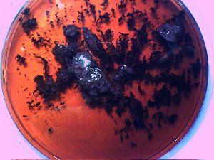
Original representative dental sample material in wine base. Essentially all individuals tested thus far produce varying degrees of this dental filament material. This is the type of material used in this test. |
|
|
|
|
|
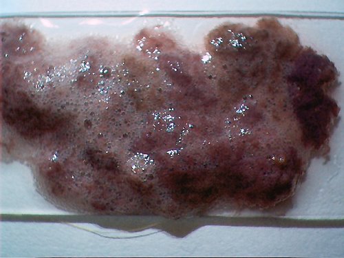
Original representative dental sample material (extracted using a wine-peroxide base) and placed onto a glass slide. The sample in this procedure has been extracted using only a wine base (no peroxide). |
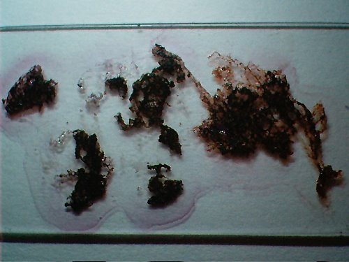
Original representative dental sample material placed onto a glass slide and dried. This dried sample is presented for comparison purposes only and is not used in this test. |
|
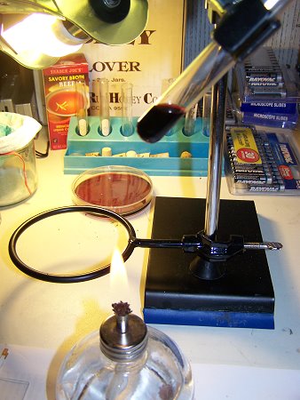
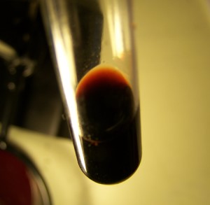
The dental filaments (from wine extraction method only – no peroxide is used for this procedure). are placed into approximately 2-3 ml. of water with one drop of a highly caustic solution (sodium hydroxide and potassium hydroxide mixture) added. Thus, a highly alkaline solution is at the core of the procedure. The exact concentration level of this solution can be determined at a later time; it does not appear to be required to be highly specific at this point. When the filaments are within the alkaline solution, an initial partial breakdown of the filaments will occur and the solution will turn darker (blackish tone) in color. The filaments do not break down in total at this point. The solution is then heated gradually and cautiously to the boiling point. |
|
In addition to the highly alkaline environment created for the filament sample, the solution is heated gradually to the boiling point as described above. This heating process appears to a critical addition to the procedure and a significant change of color will then occur. The solution will turn to a dark red color. The reddish tint that develops can be seen at the upper portion of the photographed solution above. The color of the solution at this point does indeed appear blood red, and visually does match that of blood in solution. It is possible that a hemoglobin or protein transformation is incited at this point, and the additional heat in combination with the caustic solution produces this final result. Specific tests for hemoglobin are inconclusive at this stage of the research, and a full protein analysis (not restricted to hemoglobin) is required at this point. This combination of heat and strong alkali solution would normally be considered to be detrimental to most biological processes. It appears that microscopic examination of the solution is facilitated by placing a high concentration of filaments within the solution. |
|
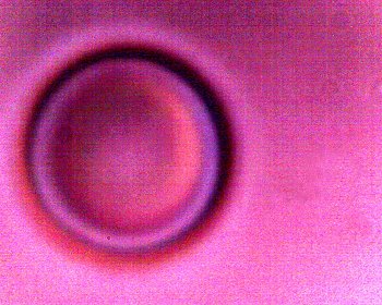
If a drop of the concentrated solution is placed upon a glass slide with a cover slip and placed under the microscope at sufficient power, numerous erythrocytic forms such as that above have been found in all cases considered. Detailed microscopic examination does indeed satisfy all visual and metric expectations of an erthrocye, or red blood cell, including biconcavity. Examination occurs over approximately a half hour interval after creating the slide. Prolonged exposure(i.e., 1day +) to this chemical environment appears to destroy all recognizable cellular forms. Please also refer to the previous report entitled “Blood Issues Intensify” of April 2009 that demonstrates the existence of blood and hemoglobin at the forensic level from cultures developed from this same dental material. Detailed protein analysis is a future requirement; such analysis cannot take place without an increased level of support and resources. Improved microscopy methods and equipment have been developed to permit viewing of the structures at this level; the magnification of this image is approximately 8000x and the structure measures approximately 6-8 microns in diameter. Conventional microscopy will peak at approximately 1000-2000x. The availability of an electron microscope would be expected to provide greater detail. There are several interesting observations that can be made of these particular erythrocytic forms, however. The first of these, as itemized above, is the extreme geometric regularity of the forms of the cells. They appear to be essentially of regular and flawless geometric form; no human blood samples examined thus far demonstrate this level of uniformity. It is this observation which asks us to consider the existence of an artificial blood form here, or at the very least the consideration of a manipulated or altered cell of some fashion. A second observation is that more variation of size (not form, however) will occur than within human samples observed. This appears to be a result of the chemical environment that allows this reconstitution process to take place. The cells will change in size during observation on the microscope stage, and some of them will reach abnormally large diameters estimated up to approximately 20-25 microns. In addition, some of the cells will reconstitute to a smaller diameter than a human cell, down to a level of approximately 4 microns in diameter. The average size of the cells appears to coincide closely with that of the human species, on the order of 6-8 microns in diameter. It does appear to be a remarkable event of discovery that this particular combination of chemical and thermal environments causes this apparent reconstitution to take place; such conditions would not be anticipated for most normal biological processes. This is another factor in the consideration of an artificial or altered biological form. It is relevant to note that previous research efforts that first uncovered dessicated erythrocytic forms also included the boiling of the solution within some of the procedures. It was at that earlier time that an understanding of hostile and adverse environmental effects upon the unique erythrocytic structures identified was reached. |
|
|
 |
|
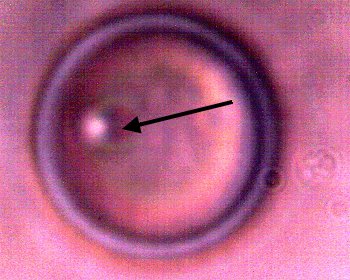
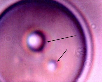
An equally important and additional observation must be considered. If the research of this site is reviewed over the past several years, it will be seen that special attention has been drawn to the existence of a sub-micron spherical structure commonly being observed within numerous human blood samples. For instance, please refer to the paper entitled “Morgellons: 5th, 6th & 7th Match“, January 2008 with special attention to the Gram stained blood cell samples as is repeated on the right side of the two images above. Further information on this particular structure has been limited by the technology available to this researcher. Further progress on this matter has long required additional resources, such as electron microscopy. This researcher has maintained a strong and particular interest in this specific structure since it was first reported. No subsequent progress on identification of this structure has been made, beyond the initial proposal that Chlamydia-like forms should be considered. This structure must be identified; further support and resources are required to accomplish this task. It is now of tremendous interest and of high importance that similar, if not identical structures, are being observed within the current reconstituted samples which are the subject of this report. The arrow on the left photograph shows such a sub-micron structure that has now identified within a dental sample that has been chemically broken down. These structures are commonly associated with the erythrocytic forms that have been discovered, both internal and external to the cells. The particular example shown also appears to be an intracellular form, as in the paper referenced above. This finding is highly suggestive that this alteration of the erythrocytic form is deliberate, and that it can produce a similar result within the general bloodstream of the human body. Again, the geometric regularity is also indicative of an artificial process that has been developed to produce this result. It also strongly indicates the likelihood of genetic transfer or manipulation in the process chain. |
|
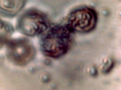
Additional examples of intracellular structures within the erythrocytic forms reconstituted from within the filament samples. |
|
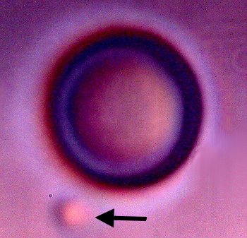
Another of many examples of geometrically smooth erythrocytic forms reconstituted from within the dental filament sample. The sub-micron structure is this example is external to the cell, as indicated by the arrow. |
This paper presents the results of further extraordinary biological observations and events that are in association with the so called “Morgellons” condition. The sample set of this report is relatively small and it must be extended. There is a remarkable consistency in the detailed observations and reports that have been made over a period of several years. This paper reaffirms the position of this researcher that blood conditions and or alterations appear to be at the crux of this situation. It is quite clear what type of work must be done to address the gravity of this situation, but additional resources must become available for this to take place. The current work now introduces the very real prospect or consideration that an artificial, or deliberately modified, process of the blood may have been introduced into the human condition. Elevated levels of research, aggressive involvement and appropriate resources must be dedicated and allocated to initiate progress on the many serious issues that have been disclosed.
Clifford E Carnicom
Aug 27 2009
Note : This paper remains subject to additional edits.
