
UNUSUAL MEDICAL FINDING
Clifford E Carnicom
Dec 07 2003
Edited Jan 30 2004
Edited Sep 05 2004
Additional Notes Sep 06 2004:
Test fibrous mass for reaction with introduction of iodine into lower gum region (blunt syringe irrigation, tincture of iodine) or affected area. Consider dark-blue chemical reaction, presence of and abundant release of insoluble fibers as a potential indicator of amyloid presence. Chemical and fibrous reaction may be significant. Mechanical removal appears to be difficult to impossible over extended period. Migration of fibrils in response to various treatments appears to be possible.
Keywords for additional research :
actinomycosis
actinomycete
fungus
iodine
amyloid
prions
mycoplasma
insoluble protein fibrils
alzheimer’s disease
degenerative tissue
proteins
starch
amyloidosis
prion diseases
Melzer’s solution
Creutzfeldt-Jakob disease (CJD)
blood test for fungus
Additional Notes Jan 30 2004:
A second fibrous mass has been retrieved from the same general location within the teeth as it has been described in the article below. The material has been retrieved from a depth of approximately 1/4 inch below the gum line with a dental needle. A remaining source of pain, although reduced, has existed since the removal of the first sample in Dec 2003. This second sample appears to be identical in nature to the original one, and it is extremely fibrous in nature. The differences between the photographs of Dec 2003 and Jan 2004 result from two main causes:
1. The use of methylene blue stain on the sample. The sample was resistant to a broad variety of stains including eosin and iodine.
2. A dramatically improved digital microscope construction, which now allows magnification to approx. 3000 without difficulty. Conventional visible light microscopy has an upper magnification limit of approximately 2000x.
The extensive fibrous nature of the sample can be seen clearly in the microphotograph immediately below. The size of the filaments measure from approximately 1 to 3 microns in thickness. For comparison, asbestos fibers are on the order of 2 microns. A human hair is on the order of 60 to 100 microns n thickness.
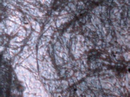
Microphotograph of second fibrous sample.
Retrieved from lower gum. Magnification approx. 3000x.
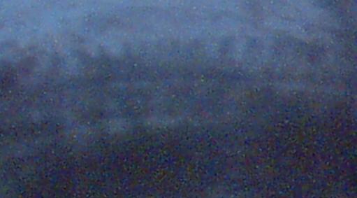
An unusual helix form has been found within the fibrous sample.
Estimated size of helix approximately 10 microns in thickness.
The traces of the helix separations are barely visible
within this microphotograph.
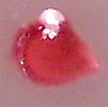
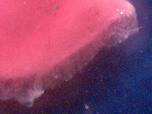
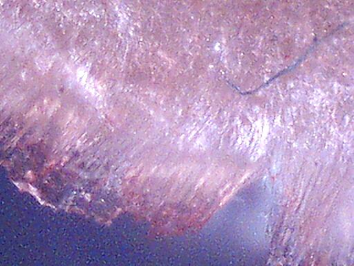
Microphotograph of removed mass : Magnification ~15x
Diameter of mass is approximately 1.5mm.
Note even circular form for a portion of the boundary at lower left.
Irregular boundary is likely affected by needle instrument used for removal.
Light spot at top is a combination of tissue and lighting conditions of microscope.
A personal dental medical situation has been under observation and examination for approximately five months. The photographs and descriptions of these findings are being made available due to the numerous sources that disclose the repeated findings of unusual microscopic fibrous material within our environment, including a direct association with the aerosol operations. In addition, a segment of the medical community has expressed an interest in identifying the origin of unusual ailments apparently associated with the presence of microscopic filaments within the skin. It is unknown whether any associations exist between the current finding and the above circumstances, but the pertinent information shall be made available for considerations.
Approximately five months ago I experienced a severe tooth ache in the second bicuspid in the lower left jaw, being the the first problem of that type in my life. I aggressively pursued the origin of the pain and found it to be most likely associated with the gum. A regimen involving the use of a water pic and a dental needle was strictly followed, and some progress on that tooth and gum soon followed. After several weeks of careful attention, the majority of pain in that particular location had alleviated. This particular tooth is unusual in that it has been described to me in early adult years as never having reached maturity, and consequently is more eroded than is usual. Ultimately, it appears that this particular tooth may have been more vulnerable to an infection or ailment, and thus may explain why the pain first appeared at this location.
In an effort to try to identify the true origin of the pain, gradual probing with the water pic and dental needle was continued beyond the immediate area for several weeks. This eventually led to the realization that the gum adjacent to several teeth of the lower left jaw had also been affected to a milder degree, with particular attention eventually between the central and left incisor of the lower jaw. The dental needle revealed a pointed source of pain deep within the gum between those two teeth. This pain was not evident unless sufficient probing to identify the source of any pain was completed. These circumstances were established approximately two months ago, and they have remained generally in a steady state until now.
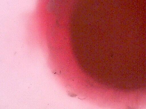
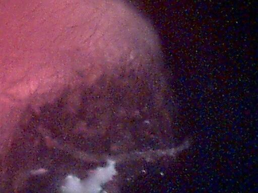
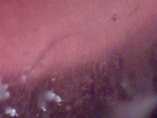
Microphotograph : Magnification 60x
Darker border of central mass visible.
Less dense and highly fibrous materials surround the central mass
The difficulty that had arisen is that this source of pain could not be eliminated, regardless of the attention and patience given over a several week to month period. No obvious degradation or deterioration in the gum adjacent to the pain origin was visible.
With continued effort, approximately one month ago, two small but visible filaments were removed from deep within the gum between these two teeth. Some time and effort were expended under the microscope to analyze those fibers. Ultimately, no conclusions of certainty could be reached on the affair, and the benefit of doubt was given to assume it was most likely of a natural origin or from the use of a toothbrush. One fiber in particular was tubular and transparent, and although the event may have been peculiar it could not be interpreted as being extraordinary.
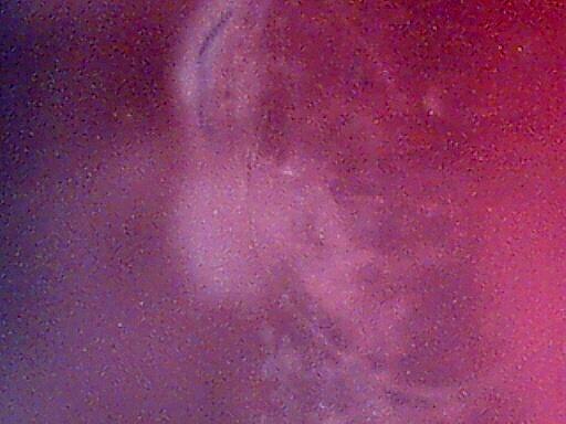
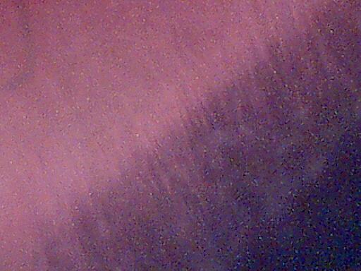
Microphotograph : Magnification 60x
Darker border of central mass visible.
Less Dense and highly fibrous materials surround the central mass.
Nature of fibrous materials slightly visible extending from border of central mass.
The findings of this day appear to be of a more unusual nature, however, and the facts of observation should be made evident. On this morning, the process of probing with the dental needle to the source of the pain continued. A visible mass eventually appeared at the base of the two teeth mentioned. The size and solid form of the material did appear to be unusual at that point, and it was subsequently collected for observation and analysis under the microscope.
The mass is approximately 1.5mm in diameter, and at first glance might appear to be of biological origin due to the reddish color. The most striking characteristic of the material, however, is that it appears to be composed primarily of filaments. The first estimate of the size of the fibers is on the order of approximately 5-10 microns in thickness. The central core of the material is rather dense, and not suitable for examination under the microscope. Various light arrangements, however, indicate that this central and generally circular mass also appears to be of a filamentous or fibrous nature. This central core remains intact at this point pending further evaluation. Several of the photographs on this page show the transition between the denser central core and the less dense boundary. It is at the boundary that numerous microscopic filaments become visible, and that they can be seen to radiate generally outward. Most of these fibers are of a transparent nature, and are difficult to photograph adequately under low power because of light characteristics and their small size. The number of fibers that can be seen appear to number on the order of thousands to scores of thousands.
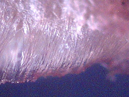
Microphotograph : Magnification 200x.
Fibrous nature of density transition zone visible.
Interspersed amongst the transparent fibers are occasional larger fibers. These fibers appear to be transparent, red or blue in color. Some evidence of these less common fibers are visible in the photographs on this page, and they are much more easily seen than the bulk of radial transparent fibers. If the bulk of the mass is found to have an natural and adequate explanation, attention will still be necessary to explain the additional larger and often colored fibers.
The level of pain of removal of the mass indicates that the material may have been integrated to a certain degree within the local nerve system.
It remains unclear at this point whether this mass of filamentous material is of biological or synthetic origin, or a combination of the two. Priority remains for a natural biological explanation of these events that have been described, but it is not available from this researcher at this point in time. Perhaps those knowledgeable in microbiology at a higher level will be forthcoming with a suitable examination and explanation.
Interspersed transparent or colored larger fibers are occasionally visible.
Colored fibers are usually blue or red. Blue and transparent fiber visible in this microphotograph.
Wet slide.Magnification 200x.
Fibrous nature more readily visible after drying of the sample.
Magnification 200x.
Fibrous nature more readily visible after drying of the sample.
Interspersed transparent or colored larger fibers are occasionally visible.
Colored fibers are usually blue or red. Blue fiber visible in this microphotograph.
Magnification 200x.
Interspersed transparent or colored larger fibers are occasionally visible.
Colored fibers are usually blue or red. Blue fiber visible in this microphotograph.
Wet slide. Magnification 200x.
Fibrous material of mass is slightly visible.
Direction of fiber orientation is generally radial. Wet slide.
Primary fibers appear to be on the order of 5-10 microns in thickness.
Central mass also appears to be fibrous in nature, but density makes observation difficult.
Magnification 200x.
Under normal dentistry conditions, it is thought that these events would likely be undetected and unrecorded.
Clifford E Carnicom
Dec 07 2003
