
CULTURE WORK IS CONFIRMED
Clifford E Carnicom
Aug 18 2008
I am not offering any medical advice or diagnosis with the presentation of this information. I am acting solely as an independent researcher providing the results of extended observation and analysis of unusual biological conditions that are evident.
A pathogenic form that appears to be directly associated with the so-called”Morgellon’s”condition has now been successfully, repeatedly and positively cultured from numerous independent dental filament samples over a protracted period of time.
The initial work that establishes the background of this report can be read from the paper entitled Culture Breakthrough (?), dated July 12, 2008. Confirmation of this result has been postponed until it became clear that the findings could be duplicated; this is now the case.
This work is important in that it provides a basis for the controlled study, observation, examination and modification of a primary pathogenic form that appears to underlie the existence of the so-called “Morgellon’s” condition. It is reiterated that the general population appears to be subject to the existence of the pathogen, regardless of whether certain skin “anomalies” are present or not.
It is unlikely that I will have the time or resources to conduct the studies that are called for. I will continue to do what I can when I can; proper resources are a serious issue at this point.
The public must now share in the responsibility for the progress(or the lack of it) that is dictated by this report.
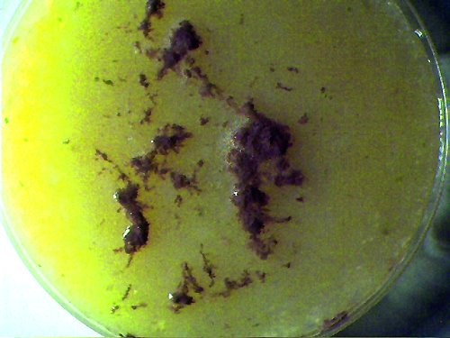
A representative original dental sample on beef-bouillon agar medium. Magnification approx. 2x.
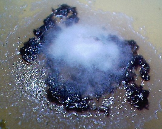
Duplicated, isolated and cultured “primary pathogenic form” growing on top of dental filament sample within bouillon agar medium.
Under high magnification, i.e., approx. 7000x, the primary pathogenic form is identical in size, shape and structure
to the expelled dental samples ( see additional photographs at high magnification below).
The time period for independent cultures to emerge from various dental samples has ranged
from a few days to a few months. The tiime of development for this propagated culture is approximately one week.
This culture form is not assured to grow on each dental sample, but has occurred thus far in at least three independent cases
over varying time intervals. A broad variety of molds, fungi and bacterial have formed on most dental samples
in addition to individual instances of the filament culture.. Magnification approx. 3x.
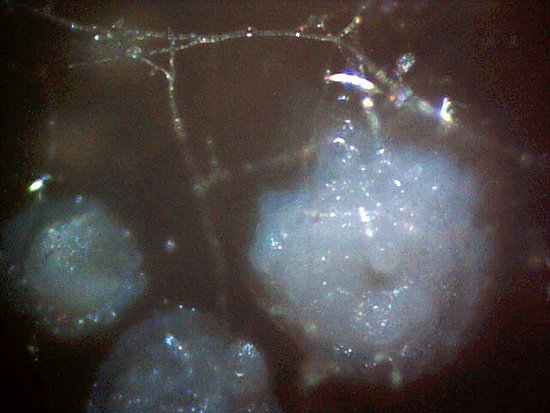
This microphotograph demonstrates one method by which the culture appears to extend its growth.
Circular colonies are often found to establish themselves on the agar medium in
a radial fashion around the primary filamentous culture. It also appears that the circular colonies
are able to withstand more adverse environmental conditions, such as a decrease in moisture.
When conditions are favorable, the filaments often form an interlocking web across and between the spherical colonies.
Individual interconnecting filaments are visible within this photograph at relatively low power.
If the conditions are highly favorable to growth, (i.e., increased moisture, nutrients and a dental sample base),
the filament culture can rapidly increase as in the first photograph of this report.
Once a filament culture has developed, it appears difficult to degrade; no such degradation
has occurred to date even if environmental conditions become more adverse.
Some cultures under study are now approximately 3 months of age.
Magnification approx. 400x.
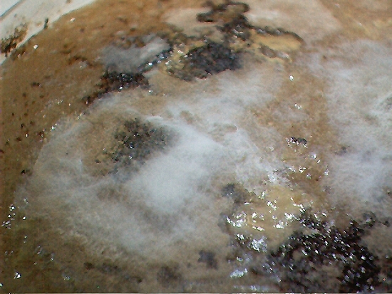
Another example of variate culture growth on the dental filament (dark regions) samples.
Bacterial and fungal forms went through several stages of evolution on this culture medium.
The culture has eventually culminated with the appearance and gradual growth of the primary pathogenic
form(white filamentous growth) after a 2-3 month period of sustained observation. Magnification approx. 2x.
The following images are excerpted from the previous paper entitled “Culture Breakthrough (?), dated July 12, 2008.
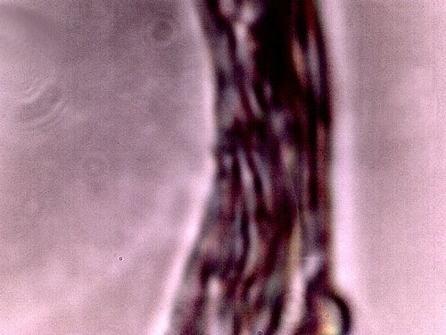
An original DENTAL filament sample under the modified microscope.
Additional dental filament sample microphotographs are
available on this site; uniformity of structure,size and form is apparent.
This is to be considered as the “primary pathogenic form”
for the purposes of this report. Magnification approx. 7000x.
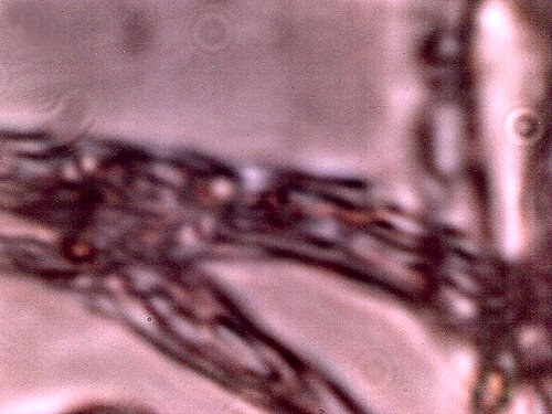
The filament from a bouillon agar culture medium growth under high magnification. It appears in all
major respects(size, structure, form) to be identical to the “primary pathogenic form.”(i.e.,the dental sample).
Encasing filament,sub-micron filament network and sub-micron oblate/spherical structures are
each identifiable within this microphotograph. This sample represents a growth on the culture medium
and it is not the original dental sample. It develops from, and as a result of the dental filament sample
and it represents a controlled development and duplication of the primary pathogenic form.
Magnification approx. 7000x.
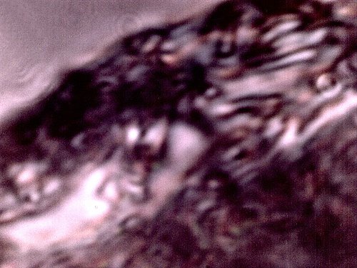
Additional microphotograph of a culture medium filament sample.
Similarity, if not identity, to the primary pathogenic form is apparent.
This pathogenic form has been identified in ALL humans that have
subjected themselves to the dental testing process.
Magnification approx. 7000x.
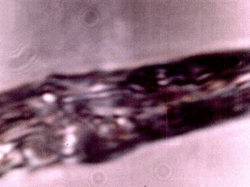
Additional microphotograph of a culture medium filament sample.
Similarity, if not identity, to the primary pathogenic form is apparent.
Magnification approx. 7000x.
Additional information will be made available if and as time and circumstances permit.
