
MORGELLONS: GROWTH CAPTURED
Clifford E Carnicom
Aug 21 2008
(High speed connection required – please allow sufficient time for loading of video)
A time lapse video under the microscope has been developed which demonstrates the cultured growth pattern and behavior of a primary pathogenic form that is in direct association with the so-called “Morgellons” condition. The general public appears to be subject to the conditions that are shown in this report.
I am not offering any medical advice or diagnosis with the presentation of this information. I am acting solely as an independent researcher providing the results of extended observation and analysis of unusual biological conditions that are evident.
(Please refer to the recent articles on this site, Culture Breakthrough (?) and Culture Work Confirmed for the prerequisites to this report).
Six hour to one minute time lapse microscope video of primary pathogenic form under culture. Magnification approx. 450x.
This video is of a cultured growth which takes place on top of a dental sample placed within an bouillon agar medium.
Please see additional images below and the recent reports on the culture work for additional information.
The time lapse video covers a period of approximately six hours and compresses the time into approximately one minute with 30 frames. The video images are time stamped in the lower right hand corner. The time interval between successive images is approximately 12 minutes. At approximately one hour into the sequence, extending filaments can be clearly seen (left center) to emerge from a primary filament. The network continues to densify from that point forward. The width of the primary filament (larger size) is approximately 12 microns in thickness, which is in accord with previous measurements for the encasing or bounding filament from direct biological samples. A reasonable estimate of the narrow filaments is on the order of sub-micron to micron range, also in accordance with previous measurements of the sub-micron internal filament network.
From the discovery shown here, it would appear that the encasing filament serves to provide feeder or extension filaments which serve to extend the growth of the pathogen. The estimated growth rate of the extension filaments on this particular culture is on the order of 50 microns per hour, or roughly the width of a thin human hair per hour.
Over the course of the six hours, it can be seen that the network becomes both dense and complex.
Developing a non-toxic method of visibly impeding this growth process should be at least one priority consideration for researchers of this topic.
The lighting varies due to surrounding reflection and refraction from the growth of the surrounding network. It also varies from the densification of the immediate network state. The depth of field for the photography is quite shallow due to the magnification, and occasionally the image requires refocusing to keep the primary filament in view. The lighting is from above and oblique.
This report continues to add valuable knowledge on the morphology, characteristics and behavior of at least some of the pathogenic forms that are strongly associated with the so-called “Morgellons” condition.
The following images are excerpted from the previous paper entitled “Culture Breakthrough (?), dated July 12, 2008
and Culture Work Confirmed, dated August 18, 2008.
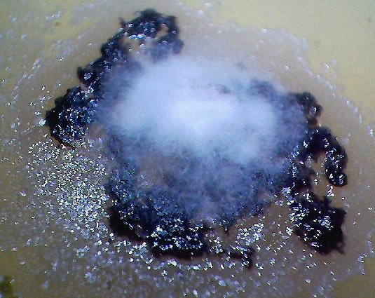
Duplicated, isolated and cultured “primary pathogenic form” growing on top of dental filament sample within bouillon agar medium.
Under high magnification, i.e., approx. 7000x, the primary pathogenic form is identical in size, shape and structure
to the expelled dental samples ( see additional photographs at high magnification below).
The time period for independent cultures to emerge from various dental samples has ranged
from a few days to a few months. The time of development for this propagated culture is approximately one week.
This culture form is not assured to grow on each dental sample, but has occurred thus far in at least three independent cases
over varying time intervals. A broad variety of molds, fungi and bacterial have formed on most dental samples
in addition to individual instances of the filament culture.. Magnification approx. 3x.
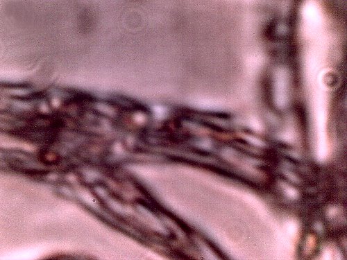
An original DENTAL filament sample under the modified microscope.
Additional dental filament sample microphotographs are
available on this site; uniformity of structure,size and form is apparent.
This is to be considered as the “primary pathogenic form”
for the purposes of this report. Magnification approx. 7000x.
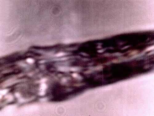
The filament from a bouillon agar culture medium growth under high magnification. It appears in all
major respects(size, structure, form) to be identical to the “primary pathogenic form.”(i.e.,the dental sample).
Encasing filament,sub-micron filament network and sub-micron oblate/spherical structures are
each identifiable within this microphotograph. This sample represents a growth on the culture medium
and it is not the original dental sample. It develops from, and as a result of the dental filament sample
and it represents a controlled development and duplication of the primary pathogenic form.
Magnification approx. 7000x.
