ERYTHROCYTES:
MAY 22
Clifford E Carnicom
May 22 2001
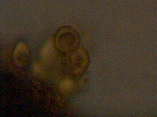
Positive visual identification of erythrocytes, or red blood cells, has again taken place from atmospheric samples collected in Santa Fe NM on May 22 2001. The method of electrostatic precipitation has again been used. Extensive counts of clusters of cells and the surrounding matrix material which readily absorbs an iodine stain were found on the microscope slides analyzed. The atmospheric samples were subjected to both sound and vapor fields to increase aggregation, in addition to exposure to the high voltage, low current electrical field.
The bi-concave surfaces, circular shapes, and dimensions of the structures again clearly identify the structures shown as erythrocytes. Measurements taken again show the size of the cells as approximately 6 microns. This represents a total of 7 out of 8 tests that have shown themselves to be a positive visual identification of erythrocyte characteristics. Both electrostatic precipitation and HEPA direct filtering techniques have been repeatedly used with identical results.
Magnification of the images shown below is approximately 5000x, obtained with the use of an oil immersion objective in combination with a digital coupler.

Magnification approx. 5000x
Bi-concavity characteristic of erythrocytes readily visible.

Magnification approx. 5000x
Bi-concavity characteristic of erythrocytes readily visible.
Clifford E Carnicom
May 22 2001


