Advances in Microscopy
Blood & Skin Filament Examinations – A Slide Show
(click on any image – controls available on each image)
Nov 19 2013
Clifford E Carnicom
A maximum magnification that combines optical and digital means has recently been achieved. The development allows, under suitable conditions and sampling, a magnification of images at a reasonable resolution up to a level of approximately 18,000 power. This method has been applied to the examination of human blood samples as they relate to the “Morgellon’s” condition. A brief introduction to the results of this recent advance in microscopy that uses relatively limited means and equipment is presented below. Relevant topics of research that arise from the study include the more detailed appearance of the bacterial-like structure that has been studied extensively by this research. The degradation of the red blood cell exterior membrane is also clearly apparent. The rather striking appearance of white blood cells, their behavior with respect to the bacterial-like component, and the internal structures that are visible within the white blood cells are of high interest. The importance of an active immune system against the bacterial-like encroachment is immediately obvious. Introductory live-blood video analysis recently performed further emphasizes the importance of the relationship of the immune system to the Morgellon’s condition. This level of awareness and visibility on the Morgellon’s condition is a direct result of these recent advances in microscopy methods and techniques. The availability of more advanced equipment, should it become available, will accelerate this discovery process.
BLOOD MICROSCOPY
[envira-gallery id=”12008″]
A brief discussion and history of the individual providing the blood control photographs above is in order. This particular individual, several years past, had blood conditions that are identical to those which are the primary subject of this paper. This individual has a history also of significant oral production of filaments (primarily in the upper oral cavity) accompanied by severe and protracted dental pain and damage to the upper teeth. Outward manifestation of skin-based filaments or skin lesions have never been significant issues with that individual.
It appears at this time that the change in the blood condition of the control individual shown above is due primarily to two main factors over a period of several years:
1. The application of the results of the extensive research results that are inherent within this site.
2. The removal of all upper teeth of the individual.
The control individual remains able to produce oral filaments, but the blood and the general health of the individual appears to have significantly improved over this same period. The chronic and severe dental pains have been eliminated at the expense of removal of the teeth. It remains of interest why the upper teeth were the primary source of injury and why they have been the primary source for oral filament production. The identification of all markers of the “Morgellons” condition and their relative importance remains a subject of much worthy discussion and research. It has long been a claim by this researcher that the state of the blood appears to be a primary factor in the evaluation of the condition along with the existence of filament forms internal to the body and their extent of distribution.
MORGELLONS SKIN FILAMENT MICROSCOPY
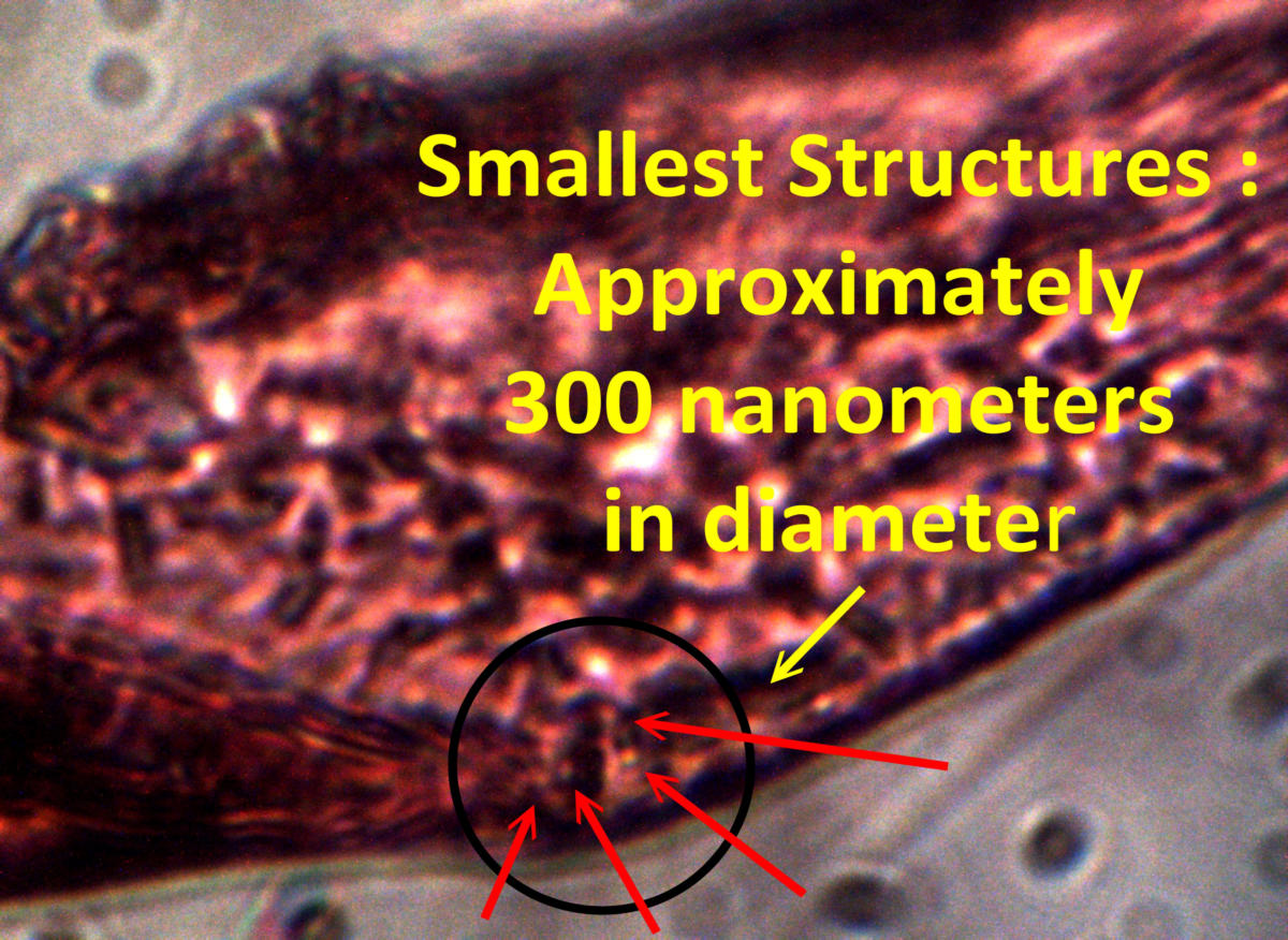
(Original magnification of all images 18,000x)
[envira-gallery id=”12009″]
The images above represent another breakthrough in the analysis of the external skin filaments that are associated with the Morgellons conditions. The majority of the images are captured using oil immersion techniques in combination with a digital-optical modified microscope. This set of images are the most detailed to date that are known and they show a plethora of internal structural forms. A more reliable measurement of the “bacterial-like” (i.e., chlamydia-like) structure has now been acquired with the use of the advanced techniques. This measurement is on the order of 300 nanometers, and thus the world of nanotechnology is now within the domain of Institute research. It is now clear that both the internal sub-filament structure and the bacterial-like forms are both on the order of 300 nanometers in diameter or width. This measurement is within the range of the larger viruses and of the smallest bacteria. A fair amount of effort is required to acquire the imagery shown.
It will be found that the shape, size, geometry, chemistry, and infra-red spectral response is identical for both the 300 nanometer structure within the blood and the 300 nanometer structure within the exterior skin filament. Ultimately it will be understood that the structure is also identical to that within the “environmental filament” so extensively studied by the Institute in the past. They are all of one and the same cloth, and at some point it will be equally understood and accepted that “Morgellons” does indeed have an environmental source for its existence.
The photos above show a great deal of detail with respect to the internal filament structure encased within the exterior filament housing, the sub-micron structures, the pleomorphism quality, aerosolization of the filaments at the filament boundary, and numerous budding and generating structures that are at the heart of its growth process. Detailed examination will show that these same forms and processes occur within the blood of those affected by the so-called “Morgellons” condition and that this conclusion can be documented and replicated in a controlled environment.
There is a wealth of discussion that could take place with the photographs shown above. There is a strong and clear lineage of research over many years that leads us to these consolidated images of yet another examination of the blood and the impact of the Morgellon’s condition upon the blood. The thesis of the blood condition as a primary indicator for the existence of the Morgellon’s condition remains. The evidence supporting the broad display of these effects by much of the general population remains in place. The means and methods may improve slowly over time, but the general conclusions of harm have been reached some time ago. My time and opportunity constraints force me to leave this extended discussion for a later date, as another paper in progress for more than a year demands its conclusion. The need for a honest and thorough investigation, the call for full disclosure, and the dedication of resources to bring about an end to this suffering remains in place.
Sincerely,
Clifford E Carnicom
(Born Clifford Bruce Stewart Jan 19 1953)


