MORGELLONS: GROWTH CAPTURED

MORGELLONS: GROWTH CAPTURED

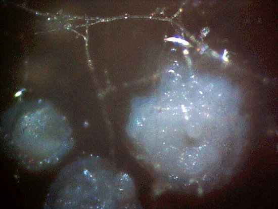
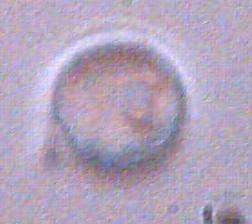
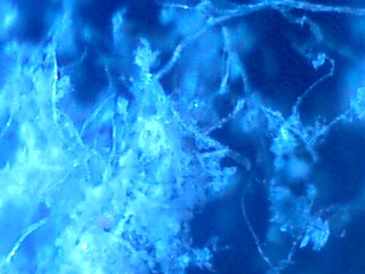
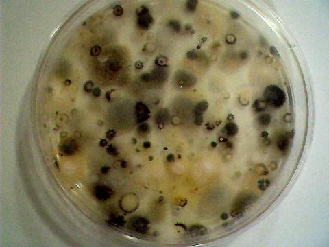
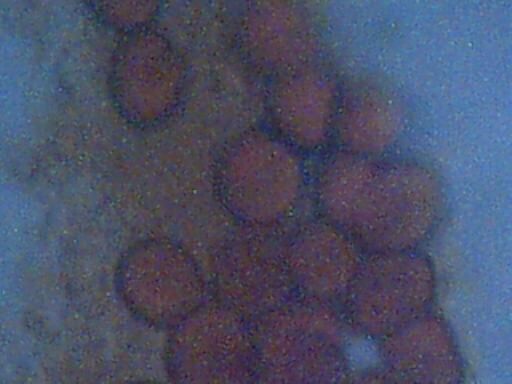
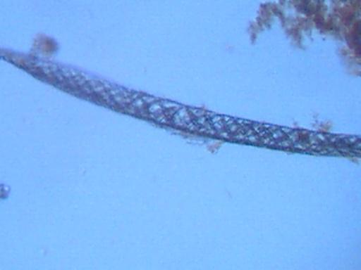
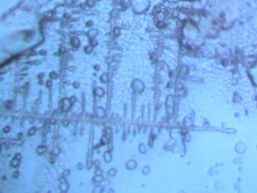
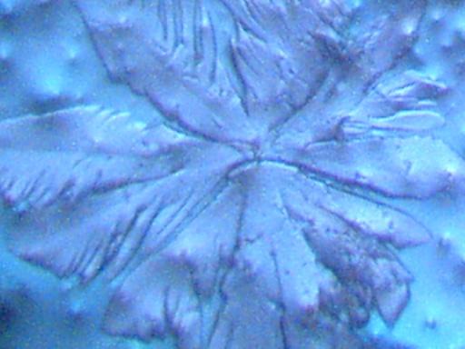
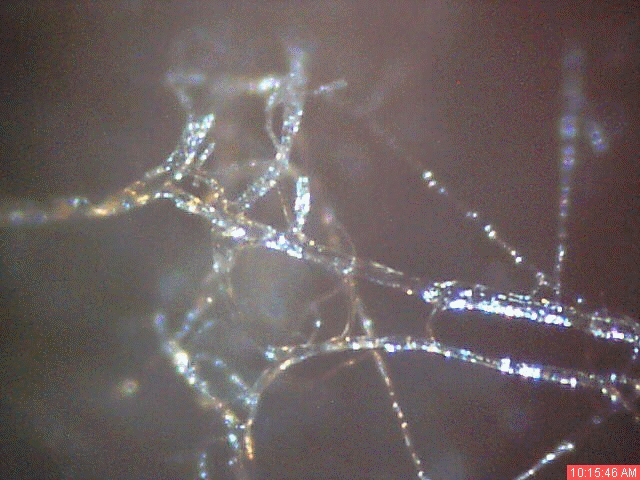
A time lapse video under the microscope has been developed and presented on this page which demonstrates the cultured growth pattern and behavior of a primary pathogenic form that is in direct association with the so-called "Morgellons" condition. The time lapse video covers a period of approximately six hours and compresses the time into approximately one minute with 30 frames. From the discovery shown here, it would appear that the encasing filament serves to provide feeder or extension filaments which serve to extend the growth of the pathogen. The estimated growth rate of the extension filaments on this particular culture is on the order of 50 microns per hour, or roughly the width of a thin human hair per hour. This report continues to add valuable knowledge on the morphology, characteristics and behavior of at least some of the pathogenic forms that are strongly associated with the so-called "Morgellons" condition.
CULTURE WORK IS CONFIRMED

CULTURE WORK IS CONFIRMED

A pathogenic form that appears to be directly associated with the so-called "Morgellon's condition” has now been successfully, repeatedly and positively cultured from numerous independent dental filament samples over a protracted period of time. Confirmation of this result had been postponed until it became clear that the findings could be duplicated; this is now the case. High magnification microscopic images are provided to the reader for further understanding of what is being presented and the implications of this work. This work is important in that it provides a basis for the controlled study, observation, examination and modification of a primary pathogenic form that appears to underlie the existence of the so-called "Morgellon's" condition. It is reiterated that the general population appears to be subject to the existence of the pathogen, regardless of whether certain skin "anomalies" are present or not. As it is unlikely that Carnicom will have the time or resources to conduct the studies that are called for going forward in this research, it needs to be known that proper resources are a serious issue at this point. The public must now share in the responsibility for the progress (or the lack of it) that is dictated by this report.
CULTURE BREAKTHROUGH(?)

CULTURE BREAKTHROUGH(?)

Work has been conducted over the past one to two months that appears to be important and it may have significant impact on current research into pathogens being found in samples discussed in the most recent Carnicom papers.  It appears as though a primary pathogenic form under evaluation that is associated with the so-called "Morgellon’s" condition may have been successfully cultured.  If this proves to be the case, it offers the potential to begin very serious research on the methods to control, inhibit, reduce or eliminate the pathogenic forms within the human body. Unknown pathogens are difficult to identify, treat and remove if they exist only within the body; there is tremendous benefit if such pathogens can be grown or developed in a culture medium under controlled conditions.  This report may offer a pathway to that process. Further research at this time continues to show identical pathogens in samples that are viewed under an enhanced microscope setup, that without such enhancement, these images and what they reveal would not be possible. This would not have allowed the progress in this area of study by Carnicom that will possibly have tremendous positive implications for all of humanity.
Instructions for Boosting Your Microscope’s Power to Examine Your Own Samples

Instructions for Boosting Your Microscope’s Power to Examine Your Own Samples

Presented in this work are instructions for building an affordable yet fairly powerful microscope imager using a microscope and a typical webcam used with computers. This setup will allow anyone to get acceptable images of samples that have been discussed in the most recent Carnicom papers regarding the pathogens being found in people blood, gums, etc. A concerned citizen wrote to Carnicom in this paper, and describes using a Logitech Quick Cam, an empty film canister, and tape to enhance the magnification of a standard microscope by the use of a webcam's CCD sensor. It is noted that there is ample concern to have as many people reproduce Carnicom's results by the use of such a setup as is described here.
UNUSUAL MEDICAL FINDING

UNUSUAL MEDICAL FINDING

After suffering from a severe toothache, fibrous material was found under the gum line between two teeth. After repeated attempts at locating the origin of the pain, a dental assistant used a dental needle and revealed a source of pain between these two teeth. Eventually two small but visible fibers were removed from this area of pain. Some time and effort were expended under the microscope to analyze those fibers. Ultimately, the benefit of doubt was given to assume it was most likely of a natural origin or from the use of a toothbrush. At a later date, a visible mass eventually appeared at the base of the two teeth mentioned. The size and solid form of the material did appear to be unusual at that point, and it was subsequently collected for observation and analysis. The mass is approximately 1.5mm in diameter, and appears to be of biological origin due to the reddish color. The most striking characteristic of the material is that it appears to be composed primarily of filaments. Most of these fibers are of a transparent nature, and are difficult to photograph adequately under low power. The fibers that can be seen appear to number on the order of thousands to scores of thousands. It remains unclear at this point whether this mass of filamentous material is of biological or synthetic origin.
EXTRAORDINARY BIOLOGICAL OBSERVATIONS

EXTRAORDINARY BIOLOGICAL OBSERVATIONS

Further microscopy analysis of biological materials collected with HEPA air filters is presented here, as a continuation of roughly five years of similar study by Clifford Carnicom. Pictures of biological growths in petri dishes after various incubation periods are attached for consideration by others in identifying these biologicals. Of note is the striking difference between the control group growth, which remains constant over time, and the cultures developed from HEPA-weak saline solution that continue to flourish.
SUB-MICRON PARTICULATES ISOLATED

SUB-MICRON PARTICULATES ISOLATED

Current research involves a process of collection of atmospheric samples with the use of a plate ionizing filter from a citizen’s HEPA filter. This material is unusual in nature and is composed of two primary forms : fairly uniform fibrous and crystalline/powder materials. This material has been collected, placed into solution and subjected to electrolysis. A definite and repeatable chemical reaction does take place, which results in the formation of a highly insoluble precipitate. The best current analysis of that material positively identifies the existence of a metallic salt. The best analysis of the nature of that metallic salt is that of a barium compound which releases positive metallic ions in solution. In addition, direct visible observation of the precipitate under extremely high magnification detects the presence of reasonably uniform spherical sub-micron particulates within the electrolysis result. The specific gravity of these particulates is greater than that of water. Pictures of these samples are attached in this work.  It has long been postulated that the size of the aerosols under examination is in the sub-micron range, and that extremely high magnification will be necessary for detection. The size range of the aerosols has previously been estimated at approximately 0.5 microns in size as a result of atmospheric light effects; this conforms to the current observations.
ATMOSPHERIC MOLD IS ABUNDANT

ATMOSPHERIC MOLD IS ABUNDANT

An atmospheric test specific to mold detection has been conducted on March 10, 2003 in rural Santa Fe, New Mexico. The results appear to indicate a sufficient cause for concern, as a large number of colonies of several species of mold (Aspergillus, Penicillum, Cladosporium, and Rhizopus) have evolved during the incubation period in a dry high desert environment during a period of extended low moisture. Several species are shown in images on this page. There is clear and direct association between the abundance of mold and respiratory, allergic and asthmatic conditions in humans. It is hoped that additional testing will be commenced by citizens across the nation and globe using indoor and outdoor samples to assess the extent and nature of this environmental condition. Those knowledgeable in mycology are requested to offer their expertise in this matter.
ERYTHROCYTES, MATRIX & MOTILE BACTERIA

ERYTHROCYTES, MATRIX & MOTILE BACTERIA

After again conducting microscopic sessions upon particulates collected from an outdoor HEPA filter in Santa Fe, NM in 2002 and finding unexpected biological components, Carnicom further asks the professional community for input to his findings. Images from this series of analysis, as well as those from earlier tests are presented, as are links to the previous research papers showing very similar results to this analysis and a description of the collection process used to collect these samples. NOTE: After asking for input from the professional community for help in analyzing these samples, a citizen on the Carnicom.com message board wrote giving input to the analysis that Carnicom has presented in this page. This person suggested that what Carnicom has been seeing in these samples is not bacteria, based on the fact that the samples were put into distilled water, and that red blood cells, when put in distilled water, will burst within seconds. The writer further claims that red blood cells are not ‘designed to live in a neutral environment’, but rather in salty environments, and that the samples look like pollen. Carnicom’s response to these claims is to seek further professional input. A main inconsistency in the citizen’s claims above include the fact that these appear to be altered (desiccated) blood cells, and such wouldn’t necessarily conform to the same reaction as normal red blood cells. Further modifications may have been applied to these cells as well, making them not susceptible to being destroyed by being placed in distilled water.
RAINWATER SAMPLES: MICROSCOPE VIEWS (II)

RAINWATER SAMPLES: MICROSCOPE VIEWS (II)

More photographs of rainwater concentrate as viewed under a microscope are presented here as a complement to investigations recently presented on the Carnicom.com website (see previous RAINWATER METALS, CRYSTAL CHEMISTRY, and RAINWATER SAMPLES: MICROSCOPE VIEWS papers). These photographs depict primarily a log of recurring structures which are found under various conditions, rather than an analysis of such structures. These structures in these microscope pictures appear to be fibers, metal oxides, and other unidentified materials. All citizens are urged to participate in the process of further collection of rainfall samples, subsequent distillation or concentration and the identification of material substances within. Any assistance provided by other researchers or sources is welcome.
RAINWATER METALS: MICROSCOPE VIEWS

RAINWATER METALS: MICROSCOPE VIEWS

Photographs of rainwater concentrate as viewed under a microscope are presented here as a complement to investigations recently presented on the Carnicom.com website. These photographs depict as evaporated crystalline form as well as a wet slide mount of the samples. The materials in these samples under microscope appear to be composed of several distinctive and complex forms, with the dominant material being a metal oxide. The presence of fibrous materials, measuring approximately 1-2 microns diameter, occurs frequently and is easily visible within the wet slide mount pictures.
CRYSTAL CHEMISTRY

CRYSTAL CHEMISTRY

Recent results from examining rainfall samples using methods of crystal chemistry are indicating substantial levels of metallic particulates within these rainwater samples. The analysis of the aerosol operations by a combination of methods repeatedly results in considerable attention being given to the elements of Group II of the periodic table (e.g. – magnesium, calcium, barium). The crystalline forms primarily found in these rainwater samples and documented with microscopic pictures is that believed to be magnesium chloride. This gives an indication of the existence of ionic magnesium within the rainfall sample. A description of the method used to create the crystalline forms shown in the attached pictures is provided here, with discussion of the polarizability and deliquessence attributes of these samples.