MORGELLONS: A FOURTH MATCH

MORGELLONS: A FOURTH MATCH

An individual with Morgellon’s symptoms has recently expelled a massive volume of fibrous material from the gums of the mouth. The fibers, upon very high magnification, reveal themselves to be identical in size, structure and form to the skin fibers that are documented in detail in earlier papers mentioned below. This now brings to four the number of samples from entirely different mediums and environments that are showing similar to identical morphology. High magnification microscope images are included in this work showing identical sub-micron filaments within a major filament structures and spherical entities within the filaments seen in previous works mentioned above.
MORGELLONS: AGENTS OF INFECTION

MORGELLONS: AGENTS OF INFECTION

This paper attempts to define a hierarchy to be followed in order for a practical approach to determining the origins and implications of the pathogenic forms that are appearing to cross airborne and biological lines, and have been observed in airborne, skin, dental and blood samples. This hierarchy can be described as follows: 1) Conventional scientific expertise should be applied to the problem of identification of these two forms. 2) Modified or unconventional biological forms or interaction are then reasonable to consider. 3) Artificial, exotic and unfamiliar technologies could be explored for any relationship to unexplainable events or circumstances. There are two forms that require immediate identification as to their physical nature, function and purpose. The first of these is a sub-micron repeating filament that is enclosed within a larger bounding filament. The sub-micron filaments can only be seen with fairly advanced microscopy; the bounding filament is visible to the naked eye in many cases. The second form is a circular, spherical or oblate structure that also is measuring at the micron to sub-micron level.
MORGELLONS: AIRBORNE, SKIN & BLOOD – A MATCH

MORGELLONS: AIRBORNE, SKIN & BLOOD – A MATCH

This paper discusses the finding that there is essentially identical form, size and structure between the airborne filament samples that have been reported on extensively over the years in connection with the aerosol operations, the morphology of at least one characteristic Morgellon's fiber and with a series of blood anomalies that have recently been documented. The three main topics and their critical points outlined with supporting graphic images in this critical paper include: 1) Morgellon's fibers and skin samples - At least one characteristic fiber form from the Morgellon's condition contains within it a rather remarkable and extensive sub-micron fibrous network. 2) Blood samples - There appears to a remarkable coincidence of form and similarity between the internal structure of the Morgellon's skin fiber and the anomalous form in the blood of the same individual. 3) Airborne fiber - The latest microphotographs, at much higher magnification than was originally available in previous Carnicom papers, of the airborne fibrous sample that was sent to the U.S. Environmental Protection Agency for identification. This now clearly implicates and questions the role and relationship of the airborne filaments to Morgellon's and the blood conditions that are currently under research.
BLOOD TESTING: LASERS, MORGELLONS & FUNGUS(?)

BLOOD TESTING: LASERS, MORGELLONS & FUNGUS(?)

This work details a study performed on five subjects’ blood samples as a comparison to each other. Only one had outward symptoms of the Morgellon’s condition – skin sores, lethargy, etc. Though each of the other four subjects didn’t exhibit these traits, they still had anomalies in their blood, which images provided in this paper show. The use of a laser is again employed in these samples to give better detail to these abnormal changes in the subjects’ blood samples. Some questions raised at this point of the Morgellon’s research include the following: What exactly is the anomalous form that is being observed? Is there any relationship between the sub-micron granular nature of the blood anomalies with the apparent similar granular nature of the Morgellon's fiber as it has been previously observed and reported? Is there any relationship between this apparent sub-micron fibrous blood anomaly and the sub-micron fibrous nature of the Morgellon's condition? Is there any relationship to the airborne fibrous samples that have been refused by the Environmental Protection Agency for identification?
MORGELLONS: MORPHOLOGY CONFIRMED

MORGELLONS: MORPHOLOGY CONFIRMED

Several different fibers from the same subject (whose story about his suffering with the Morgellon's condition is included in this paper) were examined in the writing of this report. Those fibers are scrutinized under varying intensities of magnification under microscopes as well as under a laser light, and the results build on a previous paper of the internal structures of the observed fibers, biological natures of the fibers, the growths, or 'budding' characteristics of the fibers and more. Attention will be called again to meet some immediate requirements for identification and further examination.
MORGELLONS: FIRST OBSERVATIONS

MORGELLONS: FIRST OBSERVATIONS

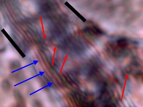
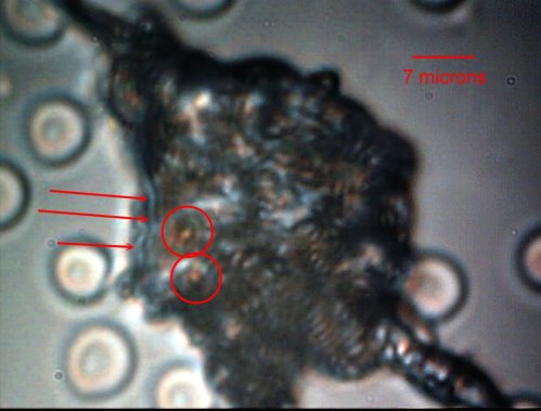
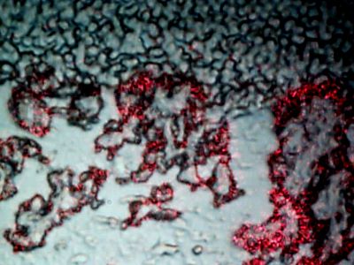
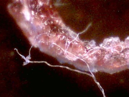
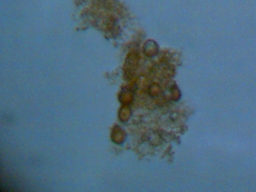
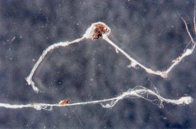
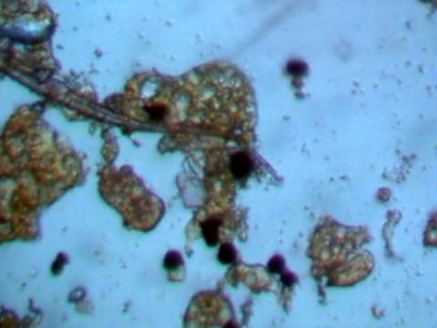
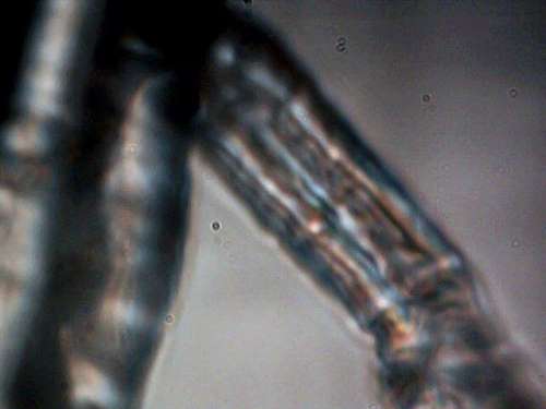
After lengthy discussions with a citizen with Morgellons, Carnicom received carefully packaged fiber samples from this person for study. The subject has provided numerous samples to a medical doctor in the past but no specific response, descriptions, photos or analysis have apparently been provided in return. One of the goals of this paper is to provide the reader with a sense of scale of these fibers, and to show a progression from the original materials as they exist from the body to the highest magnification possible with Carnicom’s equipment. Initial photographs of the filaments show little detail, but are shown also with control photographs of hair and blood cells to give a sense of scale and measurement. Further higher power microscope images show that the fibers have a much more complicated internal structure than was discernible at low magnification. These additional images appear to show innumerable sub-fibers inside the major filament. Of particular concern is that in one of the photographs of greater translucency, internal, much smaller structures of elliptical form exist. This begins to strongly suggest a biological nature to the fibers. The last major discovery by observation at this point is what appears to a "budding" structure of some sort. These structures contain two further components within. The first of these are spherical or elliptical structures at the micron level within an encasing, translucent shell. In addition, innumerable fibers at the sub-micron level emerge from the budding structure.
APR 08 2001 : BIOLOGICALS REAFFIRMED

APR 08 2001 : BIOLOGICALS REAFFIRMED

A fifth HEPA filter atmospheric sample taken in Santa Fe, New Mexico on April 8, 2001 demonstrates again the abundant presence of biological components processed by electrostatic precipitation. The biological components appear to satisfy the visual properties of erythrocytes (red blood cells). The images of these biologicals are presented here. A public appeal remains open for the professional independent evaluation of these materials being identified within atmospheric samples from different geographic regions. There exists an ethical and moral responsibility to the general public for positive identification and testing of the materials which are being found.
MAR 26 2001 HEPA: CELLS NOT FOUND

MAR 26 2001 HEPA: CELLS NOT FOUND

A third HEPA filter sample from Santa Fe, New Mexico was analyzed under a microscope with no red blood cells being found. This filter sample was collected 10 feet above ground for a duration of 10 days. These results are in stark contrast to the samples analyzed in February 25 and March 16, 2001. There remains abundant particulate and potential organic matter which requires further identification in all samples that have been acquired. These results demonstrate the need for continuous monitoring of the atmosphere at the microscopic level to ascertain the presence (or lack thereof) of certain biological components. The need for professional and independent medical, biological and chemical analysis of any HEPA atmospheric filter samples remains constant.
HEPA BIOLOGICALS CONFIRMED

HEPA BIOLOGICALS CONFIRMED

Testing with a standard HEPA air filter revealed the existence of biological components in the atmosphere. The air filter was placed 10 feet off the ground, within a non-agricultural, non-industrial, high desert rural area, and the exposure time for the test was one week. The results of this work indicate biological components which again satisfy all the visual characteristics of red blood cells in air samples taken. The methods used in this testing of filtering and identification are now available to all citizens with modest means. The identification of the cell types found, and an accountability for their existence is of critical importance to the public welfare, and it is hoped that independent researchers, professionals and activists will contribute to that cause.
ADDITIONAL FIBER PHOTOS RECEIVED

ADDITIONAL FIBER PHOTOS RECEIVED

Additional fiber samples were sent to Carnicom that were found in Diamond Springs, California, Rancho Murietta, California, and Folsom, California. The material in these samples appears to be identical to that analyzed and documented previously. These samples also appear to be identical to those samples sent to the United States Environmental Protection Agency (EPA), the agency that refuses to identify the material in the samples. Photos of these samples are shown in this paper.
GEL COMPONENTS

GEL COMPONENTS

A gel sample, identical to two others previously identified, is analyzed in this paper. This sample was sent in by a witness who found numerous identical samples in her yard after small planes and helicopters flew over her house just before the samples were found. There appears to be a clear cellular structure in the sample that absorb iodine stain readily and become darkened in color. Reports of serious ill health have been associated with this gel material.