
The Source of Blood Coagulation: The Cross Domain Bacteria (CDB)
Aug 26 2023
Clifford E Carnicom
The case can now be made that the primary mechanism causing the widespread increased coagulation of blood in the “Covid Era” is the Cross Domain Bacteria (CDB). This biological entity, synthetic or otherwise, is the subject of extensive inquiry at Carnicom Institute(CI) for many years.
This conclusion can be reached with the suitable use of microscopy alone. This paper will depend primarily on imagery to make the case, but it will also include some of the historical framework that helps to interpret the coagulation problem within the context of the Covid Era. The change and increase in coagulation/clotting appears to result from an apparent attractive force (i.e., electromagnetic, chemical, etc.) induced, aggravated or enhanced within blood that does show itself to correlate with the advent of the Covid Era.
There are two methods of microscopy that will be of complement to one another. The first is that of a traditional dried blood smear(assuming the method remains viable), and the second is with the use of live blood microscopy. In one case, the blood is static and dry. In the other case, the blood is fluid (at least initially). The historical impact, of the CDB upon blood, observed and documented now over decades, is an important aspect of the understanding before us. Undoubtedly, the Covid Era added another dimension to the earlier problems recorded, and this will also be discussed within this paper.
I will provide an image early in this paper so that the reader may have an intuitive perception as to where this discussion is leading. It will take some time to establish an understanding of what follows below. I leave it to the reader whether they wish to engage at the level required, but all of us may immediately have a sense of how remarkable but problematic the image below is.
Blood Cell Coagulation Mechanism with CDB
Original Magnification ~8000x
I hope that you will bear with me, however, as our future as a species most likely does depend upon our “engaging”.
We must start with what motivated this examination to begin with, and when it arose and why.
In August through October of 2022 a six-part research paper series was written that spans roughly a year and a half of work. The paper set deals primarily with extraordinary findings that involve the application of electrical energy to blood (unfortunately, the end result is lethal). Beyond the broader perspective afforded by those papers, the opening paper of the series is especially relevant here as it reports on unusual coagulation behavior of blood. So much so that making proper slides for the viewing of blood was difficult if not impossible. The paper series is available here.
The following statement was made in the summary at the opening of this first paper, entitled “Blood Aterations I : Coagulation“:
| “The coagulation factors appear to associate with the presence and effects of the “cross-domain” bacteria (CDB); a unique microbial life form identified and studied by Carnicom Institute over the last 25 years.” |
One might question how that perception was reached at that stage of research, but suffice it to say that it came from the changes in blood slide preparation and blood observations at the time this paper was written. Proper dried blood slides could no longer be made. The primary change over the preceding three years was the entry of the Covid Era. The essential problems with blood remained squarely resulting from the CDB, as they had for twenty years prior. but something new was now on the horizon. This change was, is, and remains centered on unknown influences from the Covid Era upon the human population.
And thus the question has remained :
What agent, force, or mechanism is responsible for causing widespread, and now devastating, changes, especially coagulation and blood clotting, in human blood on a global scale?
Generalized answers, presumptions, conjectures, misinformation, discord, speculation, sensationalism, and plenty of maybe’s are all woefully insufficient. There are plenty of all of these to surpass our all needs. When combined with the censorship and restrictions in place to access truthful information, we have generally been left to wallow in a state of confusion for the last 3 years or more (actually, more than 30). It is an absolute abhorrence at this stage that the SPECIFIC and DETAILED contents of mass worldwide injections to the population remain unknown and are perpetual fodder for the chaos just mentioned. This is not by accident, but by design.
Unfortunately, the resources of CI have been relatively minimal, for a number of reasons, over this same time period. And as Stevie Nicks sings, “I’m getting older too…” Nevertheless, a chink in that armor has been opened with recently regained access to sufficient microscopy.
And so now we need to start looking at some images.
As mentioned, both a dried blood smear and a live blood capture can be used to complement one another in this inquiry. All photographs on this page come from the same individual (no Covid “vaccination”) at the same point in time. Let’s begin with one photo from each method:
Static Dried Blood Smear – Selection 1 – Original Magnification 3200x
Live Blood – Selection 2 – Original Magnification 3200x
These photos clearly demonstrate the need for a comprehensive examination of blood under different circumstances. It may be difficult to accept that both photos come from the same individual at the same time, but this will be the case for all photos shown. What differs here are the conditions and method of view. As will be seen, there is much that can be learned from a more thorough study of the blood under various conditions and even location within the sample area.
Let us speak about the differences in this first photo set in more detail. Given the state of serious and drastic degradation of blood by the CDB that has been chronicled by CI over the years, the first photograph might be regarded as somewhat “normal”. The cell membranes have decent integrity, the cells have fairly even geometry, and the cells are distributed evenly across the sample region. But as will unfold, this blood is anything but “normal”, and the clue to that awareness is the widespread presence of the CDB within the blood, and those same CDB on surrounding the perimeter of the majority of cell membranes. There is a bit of a war taking place here. But in a superficial sense, we can start this paper by saying it has a mildly conventional appearance of blood, at least relative to what can be seen if we were to open this discussion further.
The second photograph shows the same blood from the same individual in the same time period, however it shows the state reached by the blood literally within seconds of being placed upon the slide in a live view mode. Suffice it say as it has been witnessed in times far past, a live blood view should be akin to throwing a bushel of ping bong balls in a lake. It is actually a marvelous thing to see the under microscope, and it is a dynamic and vibrant view of the marvel of healthy human blood. Unfortunately this is no longer as easy or as common to see. What we see is total and complete congregation (rouleaux) of blood on the slide immediately upon being placed upon the slide with a cover slip; this is far, far and everything away from “normal”. It is most certainly a dangerous situation, and serves as a harbinger to the widespread clotting of blood that is now reported in open scientific and health communities. It relates directly to the increased mortality that is now before us.
Now it is sensible to ask, how can this be? Why would two blood samples from the same individual at the same time look and behave so differently from one another, even within a matter of seconds?
The answer is to be found from a broader as well as a closer study of variation of cells upon the slides. Let’s begin the progression.
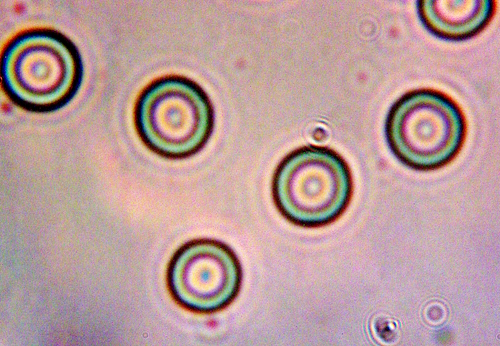
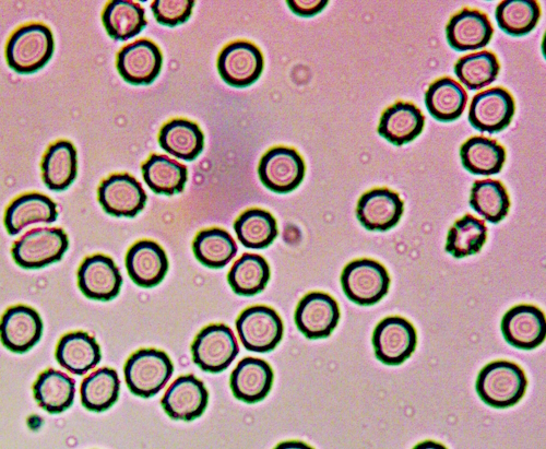
Blood Cell Microphotograph – Static Slide – Selection 3
Central CDB visible in cells (not “bulls eye” blood condition)
Original Magnification 8000x
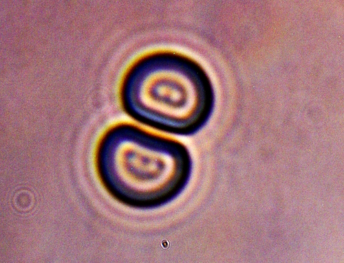
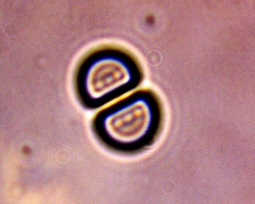
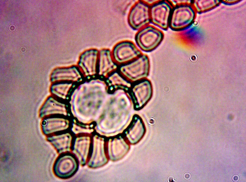
Blood Cell Microphotograph – Live Blood Slide – Selection 4
Double CDB Visible in Cells
Original Magnification 8000x
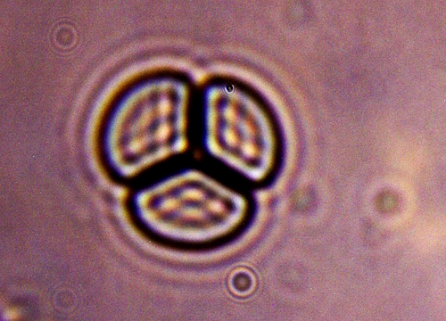
Blood Cell Microphotograph – Live Blood Slide – Selection 5
CDB Multiple Grouping Visible in Each Cell
Original Magnification 8000x
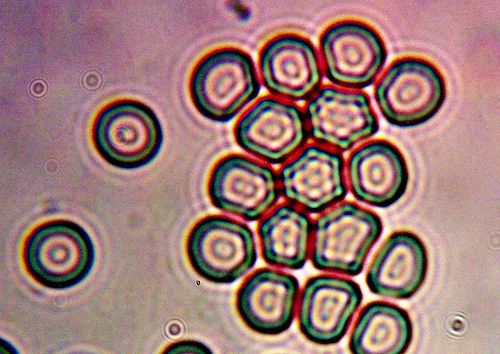
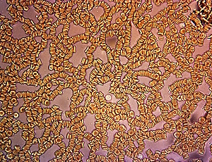
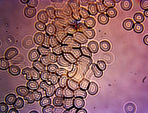
Blood Cell Microphotograph – Static Slide – Selection 6
Early Stages of Blood Coagulation
Increases Directly and Proportional to Abundance of CDB Intrusion
Notice Isolated Cells to Left with Singular Internal CDB
Original Magnification 8000x
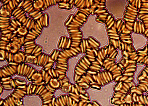
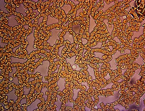
Blood Cell Microphotograph – Static Slide – Selection 7
Progressed Stage of Blood Coagulation
Directly Proportional to Severe CDB Intrusion
Original Magnification 8000x
To broaden the perspective even further, here are a few images that depict additional severe consequences of CDB impact:
Blood Cell Microphotograph – Static Slide – Selection 8
White Blood Cell Infused with CDB Attempting to Serve Immune Functions
Overwhelmed by CDB
Original Magnification 8000x
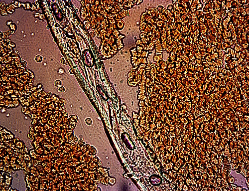
Blood Cell Microphotograph – Live Blood Slide – Selection 9
Complete Rouleaux Taking Place Essentially Instantaneously
During Preparation of Live Blood Slide Preparation
Original Magnification 8000x
Blood Cell Microphotograph – Live Blood Slide – Selection 10
Classic Example of CDB Filament within Extensive Rouleaux Formation
Original Magnification 8000x
One would think that these photographs would spawn a great deal of discussion. Even further, one might expect the top minds in the world to immediately, collectively and aggressively put themselves to work. Based on our performance as a species as a whole, this can not be assured or expected. We are behind the curve seeking three decades now, and our future as a species must be admitted to be under threat or siege. The photographs shown are not an aberration, they are representative of the state of affairs.
I do think the focus and priority of required effort before us is quite clear, as it has been. The appeals are embodied throughout the entire history of research at CI over these same decades. Carnicom Institute will proceed to the best of its capability but that history is inevitably finite. Godspeed, as it is said.
Clifford E Carnicom
Aug 26 2023
Born Clifford Bruce Stewart, Jan 19 1953.
